. 2002 Dec 15;30(24):5376-81.
doi: 10.1093/nar/gkf697.

A hairpin near the 5' end stabilises the DNA gyrase mRNA in Mycobacterium smegmatis

Affiliations

A hairpin near the 5' end stabilises the DNA gyrase mRNA in Mycobacterium smegmatis

Shyam Unniraman et al. Nucleic Acids Res. .

Abstract

RNA is amongst the most labile macromolecules present in the cells. The steady-state levels of mRNA are regulated both at the stages of synthesis and degradation. Recent work in Escherichia coli suggests that controlling the rate of degradation is as important as the process of synthesis. The stability of mRNA is probably more important in slow- growing organisms like mycobacteria. Here, we present our analysis of the cis elements that determine the stability of the DNA gyrase message in Mycobacterium smegmatis. The message appears to be stabilised by a structure close to its 5' end. The effect is especially pronounced in a nutrient-depleted state. These results largely parallel the model proposed in E.coli for mRNA degradation/ stability with subtle differences. Furthermore, these results suggest that the slow-growing organisms might use stable mRNAs as a method to reduce the load of transcription on the cell.


Figures
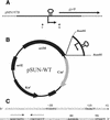
Figure 1
Figure 1
Templates used in the study. (A) The parent construct pMN197B contains part of M.smegmatis gyrB and 1.5 kb of the upstream region cloned in pUC19. The position of the transcription start site and the CHPS is indicated. The primers used for amplifying a minimal promoter region along with the 5′ untranslated region are indicated (F,R). (B) The construct used for mutagenesis, pSUN-WT. It is derived from pSD7, it contains a promoterless CAT gene (Cmr), a kanamycin resistance gene (Knr), origins of replication for mycobacteria (OriM) and E.coli (OriE), and three transcription terminators (black boxes) to prevent read-through. (C) The sequence around the transcriptional start site. The transcription start site, putative promoter elements, the dyad symmetric CHPS, the Shine–Dalgarno sequence (SD) and the protein coding region, translational start (TS), are highlighted.
Figure 2
Figure 2
Mutants obtained by random as well as site-directed mutagenesis. The three single mutations obtained in the random screen and the mutations that were engineered to disrupt and regenerate the CHPS are shown along with their effect on CAT activity relative to the wild-type activity.
Figure 3
Figure 3
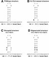
Disruption of the hairpin structure. Idealised structures of the wild-type (A), mutant from the random screen (B), site-directed disruption (C) and regenerated (D) forms of the CHPS are shown. ΔG was calculated using the web interface of the mfold algorithm as described in the Materials and Methods.
Figure 4
Figure 4
Effect of the disruption of the structure on the steady-state levels of the transcript. Primer extension analysis was performed on RNA isolated from exponentially growing cells harbouring different constructs. The wild-type, disrupted and regenerated structures are shown in Figure 3A, C and D, respectively. Representative results are shown with cultures grown in YK (A) or starvation medium (B). The average of three independent experiments is depicted graphically.
Figure 5
Figure 5
Effect of the disruption of the structure on the half-life of the transcript. RNA was isolated after exponentially growing cells harbouring different constructs were treated with rifampicin (50 µg/ml) for indicated time intervals. Primer extension was performed to quantify the level of transcript at each time point. Representative results with different cultures are shown. The wild-type, disrupted and regenerated structures are shown in Figure 3A, C and D, respectively. The average of three independent experiments is depicted graphically.

Similar articles

Cited by

References

    1. Rauhut R. and Klug,G. (1999) mRNA degradation in bacteria. FEMS Microbiol. Rev., 23, 353–370. - PubMed
    1. Regnier P. and Arraiano,C.M. (2000) Degradation of mRNA in bacteria: emergence of ubiquitous features. Bioessays, 22, 235–244. - PubMed
    1. Grunberg-Manago M. (1999) Messenger RNA stability and its role in control of gene expression in bacteria and phages. Annu. Rev. Genet., 33, 193–227. - PubMed
    1. Deutscher M.P. and Li,Z. (2001) Exoribonucleases and their multiple roles in RNA metabolism. Prog. Nucleic Acid Res. Mol. Biol., 66, 67–105. - PubMed
    1. Miczak A., Kaberdin,V.R., Wei,C.L. and Lin-Chao,S. (1996) Proteins associated with RNase E in a multicomponent ribonucleolytic complex. Proc. Natl Acad. Sci. USA, 93, 3865–3869. - PMC - PubMed

MeSH terms