Review
. 2002 Dec;70(9-10):537-46.
doi: 10.1046/j.1432-0436.2002.700907.x.

The organizing principle: microenvironmental influences in the normal and malignant breast

Affiliations
Review

The organizing principle: microenvironmental influences in the normal and malignant breast

Mina J Bissell et al. Differentiation. 2002 Dec.

Abstract

The current paradigm for cancer initiation and progression rests on the groundbreaking discoveries of oncogenes and tumor suppressor genes. This framework has revealed much about the role of genetic alterations in the underlying signaling pathways central to normal cellular function and to tumor progression. However, it is clear that single gene theories or even sequential acquisition of mutations underestimate the nature of the genetic and epigenetic changes in tumors, and do not account for the observation that many cancer susceptibility genes (e.g. BRCA1, APC) show a high degree of tissue specificity in their association with neoplastic transformation. Therefore, the cellular and tissue context itself must confer additional and crucial information necessary for mutated genes to exert their influence. A considerable body of evidence now shows that cell-cell and cell-extracellular matrix (ECM) interactions are essential organizing principles that help define the nature of the tissue context, and play a crucial role in regulating homeostasis and tissue specificity. How this context determines functional integrity, and how its loss can lead to malignancy, appears to have much to do with tissue structure and polarity.


Figures
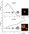
Fig. 1
Fig. 1
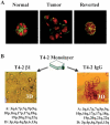
The 3D assay. Normal mammary epithelial cells, cultured in 3D gels of laminin-rich basement membrane (lrBM), arrest growth and organize into polar, acini-like structures (top). By contrast, mammary tumor cells continue to proliferate into disorganized masses (bottom). (Reproduced with modifications by permission of Cancer Biology from Weaver et al., 1995.)
Fig. 2
Fig. 2
Myoepithelial cells (MEP) provide the organizational context to luminal epithelial cells. A Luminal cells form normal acini in lrBM, but inside-out acini in collagen I (coll I), and this inverse polarization is reversed by addition of human myoepithelial cells (MEP). a, a’, a”: apical marker sialomucin (red) and basal marker epithelial-specific antigen (green); b, b’, b”: occludin (green), which orients on the apical side of the cell–cell junctions, and sialomucin (red); c, c’, c”: nuclear stain (red) and cell-ECM receptor (and basal marker) β4-integrin (green). B Cancer-derived myoepithelial cells lack the expression of laminin-α1 chain, which is a component of laminin-1, but the same cells maintain expression of laminin-α3 and laminin-α5, components of laminin-5 and laminin-10/11, respectively. (Reproduced with modifications by permission of Journal of Cell Science from Gudjonsson et al., 2002.)
Fig. 3
Fig. 3
Reversion of HMT-3522 T4–2 cells. A Reversion by inhibition of EGFR using function-blocking antibodies. Green, actin; red, nuclei. B Phenotypic reversion by inhibition of β1 integrins occurs against a constant genetic background (A reproduced from Wang et al., 1998; B reproduced with modifications by permission of Journal of Cell Biology from Weaver et al., 1997; CGH data, Joe Gray and Bissell laboratories, manuscript in preparation).
Fig. 4
Fig. 4
Inhibiting β1 integrin together with PI3K in 3D lrBM can revert or kill aggressive breast cancer cell lines. MCF-7, MDA-MB-231, and Hs578T cells were cultured in 3D lrBM either without inhibitor (control), with LY294002 (LY), an inhibitor of PI3–kinase, with AIIB2, an inhibitor of β1 integrins (AIIB2), or with both. (Reproduced with permission of Journal of National Cancer Institute from Wang et al, 2002.)
Fig. 5
Fig. 5
β1 integrin and EGFR protein levels and signal activation are coordinately modulated in HMT-3522 cells cultured in 3D lrBM but not in 2D. When 3D T4–2 cells are treated with functional inhibitors of either β1 integrin or EGFR (T4–2 treated) to revert the cells, endogenous β1 and EGFR protein levels down-modulate coordinately. This phenomenon does not occur in 2D. (Reproduced with permission of Cancer Research from Bissell et al, 1999.)
Fig. 6
Fig. 6
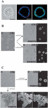
Selective alteration of signaling pathways disrupts organization of 3D structures. A MCF10A cells form growth-arrested, polarized acinar structures in 3D lrBM. a, phase contrast; b, collagen IV (red)/nuclei (blue); c, beta-catenin (green)/nuclei (blue). Scale bars: 50 mm. B MCF-10A cells in which ErbB1/EGFR could be autoactivated by addition of AP1510 show no alteration in acinar structure (b) relative to untreated cultures (a). C MCF-10A cells in which ErbB2 could be activated by addition of AP1510 showed loss of polarized organization and developed multi-acinar structures with filled lumina indicating that the combination of loss of growth and loss of apoptotic regulation creates ductal carcinoma in situ (DCIS)-like structures. (Reproduced with modifications by permission of Nature Cell Biology from Muthuswamy et al., 2002.)
Fig. 7
Fig. 7
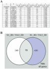
Gene expression differences between tumorigenic and nontumorigenic cell lines are influenced by 3D lrBM. HMT-3522 cell lines S1 (nontumorigenic) and T4–2 (tumorigenic) were compared for global gene expression, when grown in 2D or in 3D lrBM, using 8k human cDNA arrays. Three cultures and four slides, including a dye swap, were scanned in per experiment. Slides were scanned in using Genepix®, and data was normalized (per slide). Genes that change significantly (t-test, P<0.05) were determined using GeneSpring®. A Table showing a list of genes that change in both 2D and 3D when nontumorigenic and tumorigenic cells are compared. A normalized ratio of > 1 represents a higher expression in tumorigenic T4–2 cells; * indicates genes that change in opposite direction in 2D vs. 3D lrBM (Rizki and Bissell, unpublished data). B Venn diagram shows a comparison of the list of genes that change significantly between S1 and T4–2 in 2D vs. in 3D.
Fig. 8
Fig. 8
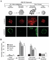
Polarized mammary structures are resistant to apoptosis induction. S1 acini within 3D lrBM were treated with E-cadherin blocking antibody to perturb polarity. T4–2 structures within 3D lrBM were treated with beta-1 integrin inhibitory antibody to restore polarity. A Confocal microscopy of beta-catenin and laminin-5. S1 and reverted T4–2 acini have junctionally organized beta-catenin and basally secreted laminin-5, in contrast to disrupted S1 and disorganized T4–2 colonies. B Apoptotic index of the cell cultures in (A), either untreated (basal), or treated with etoposide, Fas-R, or TNF-α. (Reproduced with modification by permission Cancer Cell from Weaver et al, 2002.)
Fig. 9
Fig. 9
Heterotypic 3D cultures recapitulate in vivo structures. A Coculture of human mammary luminal and myoepithelial cells in collagen I (left), compared to histologic section of normal human breast tissue (right). Green is a myoepithelial marker Thy-1; red is nuclear staining (reproduced with modifications by permission of Journal of Clinical Investigation from Gudjonsson et al., 2002). B Coculture of human luminal epithelial and stromal fibroblast cells in collagen I (left), compared to histologic section of human breast cancer (right). Staining for vimentin and counterstaining of nuclei with hematoxylin (reproduced with modifications by permission of Journal of Cell Science from Ronnov-Jessen, 1995).

Similar articles

Cited by

References

    1. Airola K, Fusenig NE. Differential stromal regulation of MMP-1 expression in benign and malignant keratinocytes. J Invest Dermatol. 2001;116:85–92. - PubMed
    1. Barcellos-Hoff MH, Aggeler J, Ram TG, Bissell MJ. Functional differentiation and alveolar morphogenesis of primary mammary cultures on reconstituted basement membrane. Development. 1989;105:223–235. - PMC - PubMed
    1. Bissell MJ. The differentiated state of normal and malignant cells or how to define a “normal” cell in culture. Int Rev Cytol. 1981;70:27–100. - PubMed
    1. Bissell MJ, Radisky D. Putting tumours in context. Nature Rev Cancer. 2001;1:46–54. - PMC - PubMed
    1. Bissell MJ, Weaver VM, Lelievre SA, Wang F, Petersen OW, Schmeichel KL. Tissue structure, nuclear organization, and gene expression in normal and malignant breast. Cancer Res. 1999;59:1757s–1763s. discussion 1763s–1764s. - PubMed

Publication types