Review
. 2002 Nov;60(5):337-53.
doi: 10.1034/j.1399-0039.2002.600501.x.

The CD1 family and T cell recognition of lipid antigens

Affiliations
Review

The CD1 family and T cell recognition of lipid antigens

Y Dutronc et al. Tissue Antigens. 2002 Nov.

Abstract

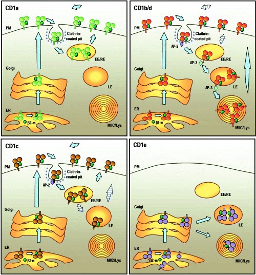
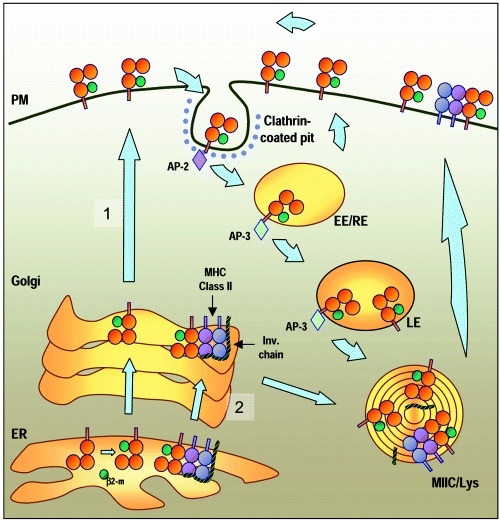
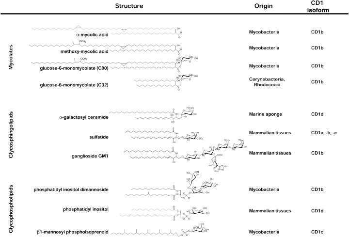
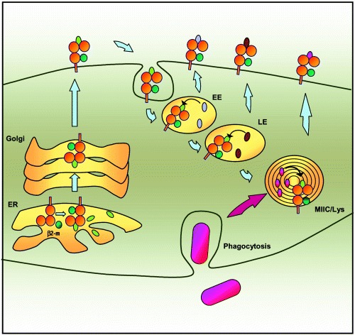
For many years it was thought that T lymphocytes recognized only peptide antigens presented by MHC class I or class II molecules. Recently, it has become clear that a wide variety of lipids and glycolipids are also targets of the T cell response. This novel form of cell-mediated immune recognition is mediated by a family of lipid binding and presenting molecules known as CD1. The CD1 proteins represent a small to moderate sized family of beta2-microglobulin-associated transmembrane proteins that are distantly related to MHC class I and class II molecules. They are conserved in most or all mammals, and control the development and function of T cell populations that participate in innate and adaptive immune responses through the recognition of self and foreign lipid antigens. Here we review the current state of our understanding of the structure and function of CD1 proteins, and the role of CD1-restricted T cell responses in the immune system.


Figures

Figure 1
Figure 1
Cellular distribution and trafficking of human CD1 molecules. Top left: CD1a. After association with β2‐microglobulin in the endoplasmic reticulum (ER), CD1a molecules are transported to the plasma membrane (PM) through the secretory pathway. CD1a spontaneously internalizes via clathrin‐coated vesicles. It is not known whether this internalization requires association with other molecules. After internalization, CD1a recycles to the plasma membrane through early/sorting and early/recycling endosomes (EE/RE). Top right: CD1b and CD1d. The cellular distributions of CD1b and CD1d molecules are very similar. After expression at the cell surface, the interactions of the intracytoplasmic tails of CD1b and CD1d with the cytosolic adaptor complex AP‐2 mediate their internalization into clathrin‐coated pits and delivery to early endosomes. From there, they access the late endosomes (LE), the lysosomes and the MHC Class II compartments (MIIC/Lys) through their interaction with another cytosolic adaptor, AP‐3. CD1d molecules undergo several rounds of recycling to the cell surface (‘striped’ arrows) and tend to accumulate in the LE and MIIC/Lys over time. CD1b and CD1d that have been loaded with lipid antigens within the endocytic system escape to the cell surface and presumably accumulate there for antigen presentation. Bottom left: CD1c. CD1c molecules transit to the cell surface through the secretory pathway and are internalized via clathrin‐coated vesicles through their interaction with AP‐2. Most of the molecules recycle to the cell surface through the EE/RE, but a small fraction can be detected in the LE. Bottom right: CD1e. Alternative splicing potentially leads to the generation of various forms of CD1e in the ER (see text). Only CD1e molecules with three lumenal α domains can associate with β2‐microglobulin (β2‐m) and leave the ER. In immature dendritic cells (DC), most CD1e molecules are present in the Golgi. After activation and maturation of DC, CD1e molecules accumulate in the late endosomes and lysosomes where they are cleaved into a soluble form.
Figure 2
Figure 2
Alternative pathways for CD1d trafficking. Most CD1d molecules are transported directly to the plasma membrane (PM) through the secretory pathway (pathway 1). There, they are internalized via clathrin‐coated vesicles and access the endosomal compartments. After recycling several times between early endosomes (EE) and the cell surface, CD1d molecules accumulate over time in the lysosomes and MIIC compartments (MIIC/Lys). From there, they eventually return to the cell surface. A fraction of CD1d molecules form a complex in the ER with MHC class II molecules and the invariant chain (Ii). These most likely enter the MIIC compartment before being expressed at the cell surface as a result of sorting at the Golgi (pathway 2). After cleavage of Ii in the MIIC, the CD1d molecules are expressed at the cell surface. At least some of these CD1d molecules remain associated with MHC class II on the plasma membrane.
Figure 3
Figure 3
Three classes of known CD1‐presented lipid antigens. The sources of the structures illustrated are stated on the right, along with the CD1 isoform(s) known to present them. Top: mycolates. These are α‐branched, β‐hydroxy fatty acids produced by bacteria belonging to the order Actinomycetales (e.g., mycobacteria, nocardia, corynebacteria and rhodococci). CD1b is known to present both free and glycosylated forms of these, including a wide variety of different mycolate structures distinguished by variations in their chain lengths, the presence or absence of double bonds or cyclopropyl groups, and the presence or absence of oxygen containing substitutions (R group) near the distal end of the longer meromycolate branch. Illustrated are a dicyclopropanated C80 mycolate lacking an R group (α‐mycolic acid), and a typical methoxy‐mycolate of M. tuberculosis. Two of the many potential forms of glucose monomycolate are illustrated, both of which are known to be presented by human CD1b. Center: glycosphingolipids. The prototype for this class is the marine sponge‐derived glycolipid (2S,3S,4R)‐1‐O‐(α‐D‐galactopyranosyl)‐N‐hexacosanoyl‐2‐amino‐1,3,4‐octadecanetriol, generally referred to as α‐galactosyl ceramide. More recent studies have demonstrated CD1‐restricted presentation of several mammalian glycosphingolipids, two of which are illustrated. Bottom: glycophospholipids. Mycobacterial forms of this class were initially demonstrated to be presented by human CD1b, including the relatively simple phosphatidylinositol dimannoside structure shown and more heavily glycosylated versions of this basic structure such as lipoarabinomannans of M. tuberculosis and M. leprae. Mammalian phosphatidylinositol and more highly glycosylated versions of this structure (glycosylphosphatidylinositols (GPI)) have been shown to bind to mouse and human CD1d proteins, although evidence for their recognition by T cells is currently limited, The fully saturated β1‐mannosyl phosphoisoprenoids isolated from M. tuberculosis and M. avium are unique at present as the only CD1‐presented antigens bearing a single alkyl chain tail.
Figure 4
Figure 4
Binding of endogenous and exogenous lipids by CD1 molecules. CD1 molecules probably bind endogenous self lipids (green ovals) such as phosphatidylinositiol and glycosylphosphatidylinositol in the ER. The association may stabilize the molecule and protect the binding groove during migration to the cell surface. After internalization from the cell surface, the various CD1 isoforms follow similar pathways into the endocytic system. They accumulate at different sites (EE vs LE vs MIIC/Lys), which allows them to collectively sample the entire endocytic network. As they pass through the various compartments CD1 molecules may exchange the endogenous lipids loaded in the ER with other lipids accumulating in the cell via fluid phase or receptor‐mediated endocytosis (lavender and brown ovals) or phagocytosis (pink ovals). Most CD1 molecules recycle several times between the plasma membrane and the endosomes, thus enhancing their chances of encountering antigenic lipids at intracellular sites.
Figure 5
Figure 5
Cellular activation events induced by αGalCer. Administration of αGalCer in mice induces a complex series of cellular events referred to as an adjuvant cascade. (1) Recognition of αGalCer through the semi‐invariant TCR activates the NK T cells and induces rapid release of IL‐4 and IFNγ. The initial activation also leads to an up‐regulation of CD40L on the NK T cells. (2) The interaction of CD40L with CD40 activates the antigen‐presenting cell (APC) which produces IL‐12. (3) The release of IL‐12 amplifies the production of IFNγ by the NK T cells and acts together with IFNγ to activate NK cells. (4) IL‐12 also plays a major role in the polarization of Th0 naive T cells into Th1 helper and cytotoxic T cells. (5) IL‐4 production by NK T cells contributes to the activation of B cells. The immunoglobulin production by B cells is influenced by the balance between IL‐4 and IFNγ. While IFNγ promotes IgG2a production, IL‐4 stimulates IgG1 and lgE secretion. (6) In addition, IL‐4 can polarize Th0 T cells toward the Th2 phenotype. Under conditions that favor the preferential production of IL‐4 following initial activation of NK T cells, this effect on Th2 polarization may predominate.

Similar articles

Cited by

References

    1. Porcelli SA, Modlin RL. The CD1 system: antigen‐presenting molecules for T cell recognition of lipids and glycolipids. Annu Rev Immunol 1999: 17: 297–329. - PubMed
    1. Calabi F, Milstein C. The molecular biology of CD1. Semin Immunol 2000: 12: 503–9. - PubMed
    1. Calabi F, Jarvis JM, Martin L, Milstein C. Two classes of CD1 genes. Eur J Immunol 1989: 19: 285–92. - PubMed
    1. Angenieux C, Salamero J, Fricker D et al. Characterization of CD1e, a third type of CD1 molecule expressed in dendritic cells. J Biol Chem 2000: 275: 37757–64. - PubMed
    1. Brossay L, Jullien D, Cardell S et al. Mouse CD1 is mainly expressed on hemopoietic‐derived cells. J Immunol 1997: 159: 1216–24. - PubMed

Publication types