Review
. 2002 Dec;6(6):500-8.
doi: 10.1186/cc1822. Epub 2002 Sep 12.

Clinical review: Myocardial depression in sepsis and septic shock

Affiliations
Review

Clinical review: Myocardial depression in sepsis and septic shock

Olivier Court et al. Crit Care. 2002 Dec.

Abstract

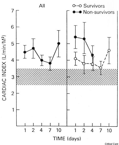
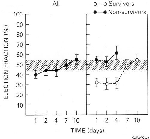
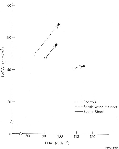
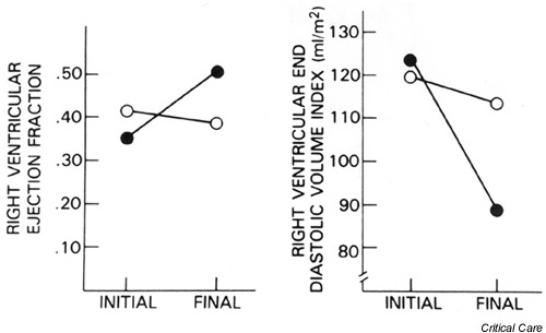
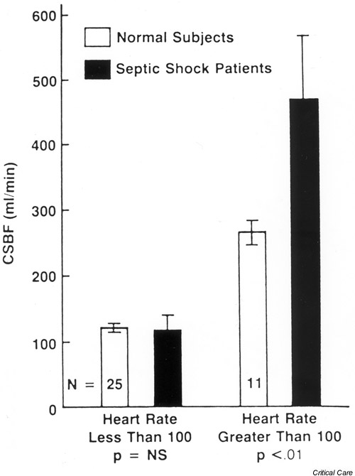
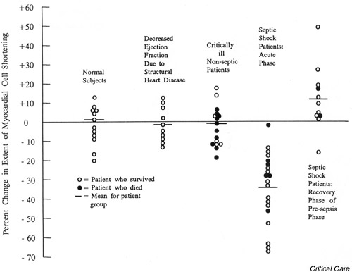
Myocardial dysfunction frequently accompanies severe sepsis and septic shock. Whereas myocardial depression was previously considered a preterminal event, it is now clear that cardiac dysfunction as evidenced by biventricular dilatation and reduced ejection fraction is present in most patients with severe sepsis and septic shock. Myocardial depression exists despite a fluid resuscitation-dependent hyperdynamic state that typically persists in septic shock patients until death or recovery. Cardiac function usually recovers within 7-10 days in survivors. Myocardial dysfunction does not appear to be due to myocardial hypoperfusion but due to circulating depressant factors, including the cytokines tumor necrosis factor alpha and IL-1beta. At a cellular level, reduced myocardial contractility seems to be induced by both nitric oxide-dependent and nitric oxide-independent mechanisms. The present paper reviews both the clinical manifestations and the molecular/cellular mechanisms of sepsis-induced myocardial depression.


Figures

Figure 1
Figure 1
The mean (± SEM) cardiac index plotted against time for all patients, survivors, and nonsurvivors. The hatched areas show the normal range. All groups maintained an elevated cardiac index throughout the study period. The difference between the survivors and nonsurvivors was not statistically significant. Reproduced with permission from [16].
Figure 2
Figure 2
The mean (± SEM) left ventricular ejection fraction (LVEF) plotted versus time for all patients, survivors, and nonsurvivors. Overall, septic shock patients showed a decreased LVEF at the time of initial assessment. This effect was due to marked early depression of LVEF among survivors that persisted for up to 4 days and returned to normal within 7–10 days. Nonsurvivors maintained LVEF in the normal range. The hatched area represents the normal range. Reproduced with permission from [16].
Figure 3
Figure 3
Frank–Starling ventricular performance relationship for each of the three patient groups. Data points plotted represent the mean prevolume and postvolume infusion values of end-diastolic volume index (EDVI) and left ventricular stroke work index (LVSWI) for each patient group. Control patients showed a normal increase of EDVI and LVSWI in response to volume infusion. The absolute increases of EDVI and LVSWI in patients with sepsis without shock were less than those of control subjects, but the slope of the curve is similar to control patients. Patients with septic shock had a greatly diminished response and showed a marked rightward and downward shift of the Frank–Starling relationship. Reproduced with permission from [17].
Figure 4
Figure 4
Serial changes in right ventricular ejection fraction and end-diastolic volume index during septic shock in humans. (a) Mean initial and final right ventricular ejection fractions for survivors (closed circles, P < 0.001) and nonsurvivors (open circles, P < 0.001). (b) Mean initial and final right ventricular end-diastolic volume index for survivors (closed circles, P < 0.05) and nonsurvivors (open circles, P = not significant). The right ventricle, similar to the left ventricle, undergoes dilation with a drop in ejection fraction with the acute onset of septic shock. In 7–10 days, right ventricular dilation and decreased ejection fraction revert to normal in survivors. Data from [26]; adapted with permission [69].
Figure 5
Figure 5
Mean coronary sinus blood flow (CSBF) in seven patients with septic shock compared with normal subjects. Flow measurements were stratified into heart rates above and below 100 beats/min. Coronary blood flow in septic shock patients equaled (heart rate <100 beats/min) or exceeded (heart rate >100 beats/min) coronary blood flow in control patients. Reproduced with permission from [32].
Figure 6
Figure 6
The effect of serum from septic shock patients and control groups on the extent of myocardial cell shortening of spontaneously beating rat heart cells in vitro. Septic shock patients during the acute phase demonstrated a statistically significant lower extent of shortening (P < 0.001) compared with any other group. Reproduced with permission from [38].

Similar articles

Cited by

References

    1. Center for Diseases Control and Prevention National Center for Health Statistics: mortality patterns – United States, 1990. Monthly Vital Stat Rep. 1993;41:5.
    1. Angus DC, Linde-Zwirble WT, Lidicker J, Clermont G, Carcillo J, Pinsky MR. Epidemiology of severe sepsis in the United States: analysis of incidence, outcome and associated costs of care. Crit Care Med. 2001;29:1303–1310. - PubMed
    1. Bone RC, Balk RA, Cerra FB, Dellinger RP, Fein AM, Knaus WA, Schein RM, Sibbald WJ. Definitions for sepsis and organ failure and guidelines for the use of innovative therapies in sepsis. Chest. 1992;101:1644–1655. - PubMed
    1. Van der Poll T, Van Deventer JH. Cytokines and anticytokines in the pathogenesis of sepsis. Infect Dis Clin North Am. 1999;13:413–426. - PubMed
    1. MacLean LD, Mulligan WG, McLean APH, Duff JH. Patterns of septic shock in man: a detailed study of 56 patients. Ann Surg. 1967;166:543–562. - PMC - PubMed

MeSH terms