. 2003 Jan 17;299(5605):411-4.
doi: 10.1126/science.1078942. Epub 2002 Dec 19.

Production of alpha 1,3-galactosyltransferase-deficient pigs

Affiliations

Production of alpha 1,3-galactosyltransferase-deficient pigs

Carol J Phelps et al. Science. .

Abstract

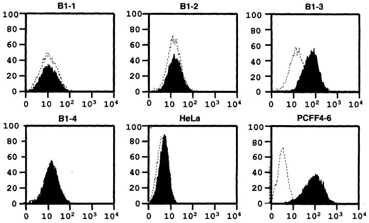
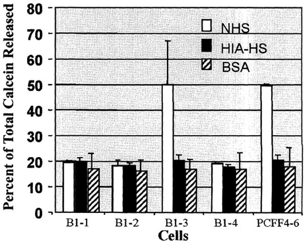
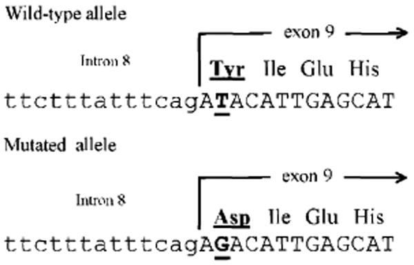
The enzyme alpha1,3-galactosyltransferase (alpha1,3GT or GGTA1) synthesizes alpha1,3-galactose (alpha1,3Gal) epitopes (Galalpha1,3Galbeta1,4GlcNAc-R), which are the major xenoantigens causing hyperacute rejection in pig-to-human xenotransplantation. Complete removal of alpha1,3Gal from pig organs is the critical step toward the success of xenotransplantation. We reported earlier the targeted disruption of one allele of the alpha1,3GT gene in cloned pigs. A selection procedure based on a bacterial toxin was used to select for cells in which the second allele of the gene was knocked out. Sequencing analysis demonstrated that knockout of the second allele of the alpha1,3GT gene was caused by a T-to-G single point mutation at the second base of exon 9, which resulted in inactivation of the alpha1,3GT protein. Four healthy alpha1,3GT double-knockout female piglets were produced by three consecutive rounds of cloning. The piglets carrying a point mutation in the alpha1,3GT gene hold significant value, as they would allow production of alpha1,3Gal-deficient pigs free of antibiotic-resistance genes and thus have the potential to make a safer product for human use.


Figures

Fig. 1
Fig. 1
Flow cytometry analysis of 680B1-1 to B1-4 cells with GS-IB4 lectin staining. Horizontal and vertical axes denote intensity of fluorescence and number of events, respectively. Dotted line represents unstained cells analyzed by a fluorescence-activated cell sorter (Becton-Dickenson, Franklin, NJ). Shadow represents cells stained with fluorescein isothiocyanate–labeled GS-I-B4 lectin (EY Laboratories, Inc., San Mateo, CA). B1-1, B1-2, B1-3, and B1-4 are fetal fibroblasts derived from four day-39 fetuses. HeLa cells, a human cell line, were used as the negative control and PCFF4-6 cells, which were the parent cells for heterozygous and DKO of the α1,3GT gene, were used as the positive control.
Fig. 2
Fig. 2
Complement lysis assay for DKO fetal fibroblasts and wild-type pig cells. Results are the average of three individual assays. Open box represents NHS. Solid box represents heat-inactivated human serum (HIA-HS) as the negative control, and hatched box represents bovine serum albumin (BSA) as the reagent control. About 20% calcein release (no cells lysed) is the base value for the negative control (HIA-HS) and the reagent control (BSA). For quality control and reproducibility purposes, we did not use fresh human serum for the assay, which usually gives about 90% calcein release. About 50% calcein release (about 40% of cells lysed) from wild-type pig cells is typical with commercial serum (frozen and lyophilized) from Sigma.
Fig. 3
Fig. 3
Sequencing analysis of the α1,3GT gene from wild-type pig cells and DKO porcine fetal fibroblasts. Upper and lower alignment show nucleotide sequence of the α1,3GT intron 8–exon 9 boundary from wild-type pigs and the second allele of the DKO pig fetuses (B1-1, B1-2, and B1-4), respectively. Small letters and capital letters denote intron and exon sequences, respectively. Underlined capital letters indicate the nucleotide where the point mutation occurred. Amino acids deduced from the correspondent mutated and wild-type DNA sequence are underlined. No other mutations were found in the coding region of the α1,3GT gene from the second allele of the DKO pig fetuses in our genomic and reverse transcriptase–PCR libraries.

Similar articles

Cited by

References

    1. Galili U, et al. J Biol Chem. 1988;263:17755. - PubMed
    1. Good AH, et al. Transplant Proc. 1992;24:559. - PubMed
    1. Cooper DK, Koren E, Oriol R. Lancet. 1993;342:682. - PubMed
    1. Galili U. Biochimie. 2001;83:557. - PubMed
    1. Sandrin MS, Mckenzie IFC. Curr Opin Immunol. 1999;11:527. - PubMed

Publication types

MeSH terms