

Review
doi: 10.1128/IAI.71.1.1-12.2003.
Molecular pathogenesis of Salmonella enterica serotype typhimurium-induced diarrhea
Affiliations
- PMID: 12496143
- PMCID: PMC143292
- DOI: 10.1128/IAI.71.1.1-12.2003
Item in Clipboard
Review
Molecular pathogenesis of Salmonella enterica serotype typhimurium-induced diarrhea
Infect Immun.
2003 Jan.
No abstract available
Figures
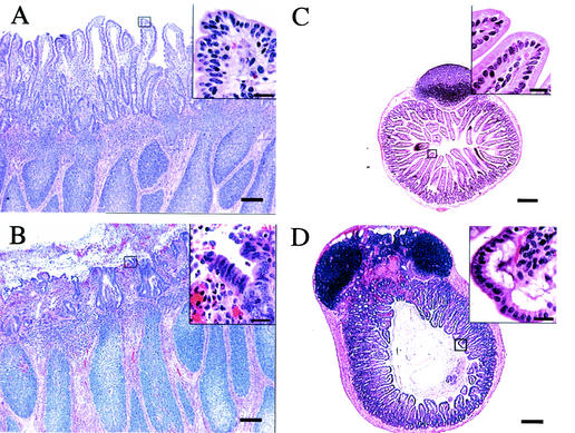
Comparative histopathology of the murine and bovine small intestine after infection of calves (A and B) or mice (C and D) with Salmonella serotype Typhimurium strain ATCC 14028. (A) Histological section of an uninfected bovine Peyer's patch (bar = 100 μm). The box covering the tip of an absorptive villus is shown as an enlargement in the insert at the top right (bar = 20 μm). (B) Histopathology of a bovine Peyer's patch at 8 h after infection of a ligated ileal loop with 109 CFU of Salmonella serotype Typhimurium (bar = 100 μm). Note the blunting of villi and the presence of a fibrino-purulent exudate in the intestinal lumen. The box covering the tip of an absorptive villus is shown as an enlargement in the insert at the top right (bar = 20 μm). Note the loss of the integrity of the intestinal epithelium. (C) Histological section of an uninfected murine Peyer's patch (bar = 100 μm). The box covering the tip of an absorptive villus is shown as an enlargement in the insert at the top right (bar = 20 μm). (D) Histopathology of a Peyer's patch from a moribund mouse at 5 days post-oral infection with 109 CFU of Salmonella serotype Typhimurium (bar = 100 μm). The box covering the tip of an absorptive villus is shown as an enlargement in the insert at the top right (bar = 20 μm). Note that the integrity of the intestinal epithelium remains intact.
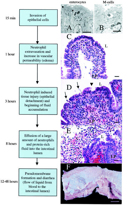
Current model of the series of events leading to an inflammatory diarrhea during Salmonella serotype Typhimurium infection of calves. (A) Transmission electron micrograph of bovine Peyer's patch at 15 min after infection of a ligated ileal loop with Salmonella serotype Typhimurium strain ATCC 14028 (109 CFU/loop). Ruffling of the brush border of an enterocyte and bacterial internalization into membrane-bound vacuoles can be seen (bar = 2.5 μm). (B) Transmission electron micrograph of bovine Peyer's patch at 15 min after infection of a ligated ileal loop with Salmonella serotype Typhimurium strain ATCC 14028 (109 CFU/loop). An M cell in the follicle-associated epithelium containing an internalized bacterium is shown (bar = 2.5 μm). (C) Focal infiltration of neutrophils in the lamina propria (LP) of an absorptive villus in bovine Peyer's patches at 1 h after infection of a ligated ileal loop with Salmonella serotype Typhimurium strain ATCC 14028 (109 CFU/loop) (bar = 20 μm). (D) Blunting of absorptive villus 3 h after infection of a ligated ileal loop with Salmonella serotype Typhimurium strain ATCC 14028 (109 CFU/loop). Note the hemorrhage and infiltration of the lamina propria with neutrophils. Arrows indicate areas where neutrophils transmigrate into the intestinal lumen (L). The arrowhead indicates the detachment of surface epithelial cells at the tip of an absorptive villus (bar = 20 μm). (E) Presence of a large number of neutrophils in the intestinal lumen (L) at 8 h postinfection of a ligated ileal loop with Salmonella serotype Typhimurium strain ATCC 14028 (109 CFU/loop). Note the hemorrhage, injury to the intestinal epithelium, and detached enterocytes (bar = 20 μm). (F) Gross pathology of the terminal ileum of a calf at 48 h after oral infection with Salmonella serotype Typhimurium strain ATCC 14028 (1010 CFU). Note the pseudomembrane formation over a bovine Peyer's patch (bar = 1 cm).
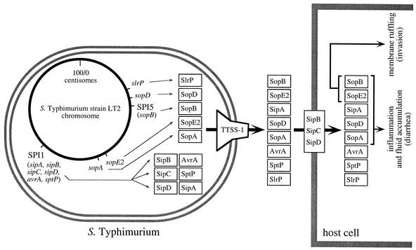
Secreted targets of the invasion-associated TTSS-1 of Salmonella serotype Typhimurium and their role in causing diarrhea in calves. The Salmonella serotype Typhimurium chromosome (circle) and the encoded TTSS-1 effector proteins (boxes) are shown in the bacterial cell (left). The TTSS-1 encoded by genes on SPI-1 forms a needle complex spanning the inner and outer membranes of Salmonella serotype Typhimurium (reviewed in reference 53) and is shown on the right side of the bacterial cell. Transport of TTSS-1 effector proteins into the host cell cytosol by the translocation complex formed by SipB, SipC, and SipD is shown on the right. Positions of genes (sopA, sopE2, slrP, and sopD) and pathogenicity islands (SPI-1 and SPI-5) on the physical map of the Salmonella serotype Typhimurium chromosome are based on the complete genome sequence of strain LT2 (63). The TTSS-1 effector genes sspH1 and sopE1 were not included in this figure since they are encoded by bacteriophages that are not present in Salmonella serotype Typhimurium strain LT2.
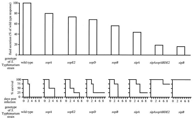
Role of TTSS-1 effector genes in causing diarrhea in calves. The relative amount of fluid accumulation in bovine ligated ileal loops elicited 8 h after infection with the Salmonella serotype Typhimurium wild type (ATCC 14028) or strains carrying mutations in TTSS-1 effector genes has been reported previously (85, 115) and is shown at the top. Mortality caused in groups of four calves after oral infection at a dose of 1010 CFU/animal with the Salmonella serotype Typhimurium wild type or strains carrying mutations in TTSS-1 effector genes has been reported previously (99, 100, 115) and is shown at the bottom.
References
-
- Ahmer, B. M., J. van Reeuwijk, P. R. Watson, T. S. Wallis, and F. Heffron. 1999. Salmonella SirA is a global regulator of genes mediating enteropathogenesis. Mol. Microbiol. 31:971-982. - PubMed
-
- Arnold, J. W., D. W. Niesel, C. R. Annable, C. B. Hess, M. Asuncion, Y. J. Cho, J. W. Peterson, and G. R. Klimpel. 1993. Tumor necrosis factor-alpha mediates the early pathology in Salmonella infection of the gastrointestinal tract. Microb. Pathog. 14:217-227. - PubMed
-
- Baggiolini, M., B. Dewald, and B. Moser. 1994. Interleukin-8 and related chemotactic cytokines—CXC and CC chemokines. Adv. Immunol. 55:97-179. - PubMed
Publication types
MeSH terms
Substances
Grants and funding
LinkOut - more resources
Full Text Sources
Other Literature Sources
Medical
