. 2003 Jan;71(1):75-85.
doi: 10.1128/IAI.71.1.75-85.2003.

Both family 1 and family 2 PspA proteins can inhibit complement deposition and confer virulence to a capsular serotype 3 strain of Streptococcus pneumoniae

Affiliations

Both family 1 and family 2 PspA proteins can inhibit complement deposition and confer virulence to a capsular serotype 3 strain of Streptococcus pneumoniae

Bing Ren et al. Infect Immun. 2003 Jan.

Abstract

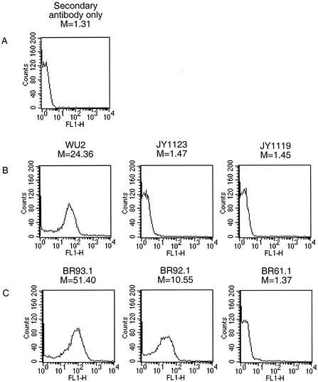
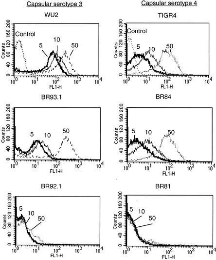
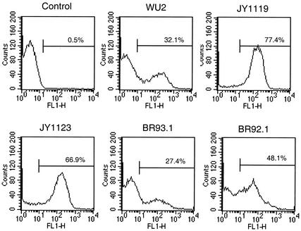
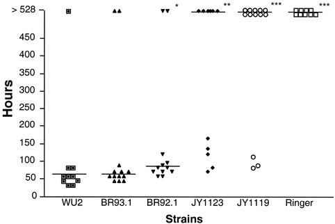
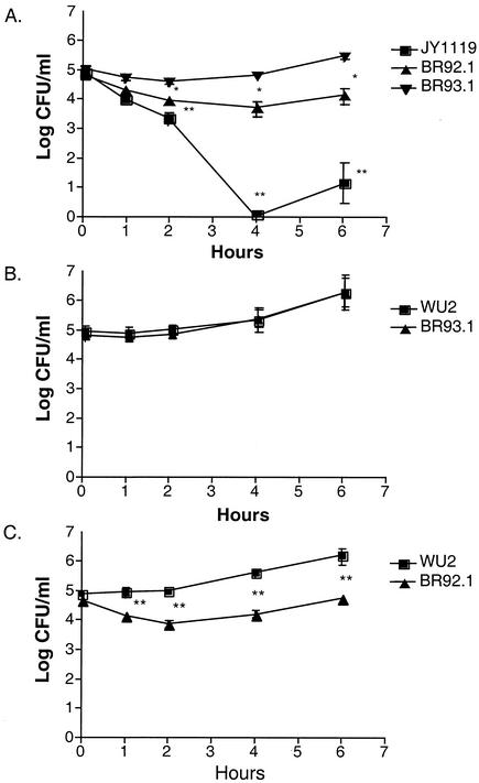
Pneumococcal surface protein A (PspA), a virulence factor of Streptococcus pneumoniae, is exceptionally diverse, being classified into two major families which are over 50% divergent by sequence analysis. A family 1 PspA from strain WU2 was previously shown to impede the clearance of pneumococci from mouse blood and to interfere with complement deposition on the bacterial surface. To determine whether a family 2 PspA can perform the same role as family 1 PspA, the family 1 PspA (from strain WU2) was replaced with a family 2 PspA (from strain TIGR4) by molecular genetic methods to make an isogenic pair of strains expressing different PspA proteins. Surface binding of lactoferrin and interference with C3 deposition by the two types of PspA proteins were determined by flow cytometry, and virulence was assessed in a mouse bacteremia model. Although the family 2 PspA appeared to bind less human lactoferrin than did the family 1 PspA, both PspA proteins could interfere with complement deposition on the pneumococcal surface and could provide full virulence in the mouse infection model. A mutant form of the family 2 PspA with a deletion within the choline-binding region was also produced. Pneumococci with this mutant PspA failed to bind human lactoferrin even though the PspA was present on the pneumococcal surface. The mutant PspA only partially interfered with complement deposition and moderately attenuated virulence. These results suggest that family 1 and family 2 PspA proteins play similar roles in virulence and that surface accessibility of PspA is important for their function.


Figures

FIG. 1.
FIG. 1.
Western blots reacted with rabbit antisera specific for family 1 or 2 PspA. Bacterial cell lysates were separated on duplicate SDS-PAGE gels and transferred onto nitrocellulose membranes. Blots were reacted with anti-family 1 PspA serum (A) or anti-family 2 PspA serum (B). Lanes: 1, WU2; 2, BR93.1; 3, BR92.1; 4, JY1123; 5, JY11119; 6, TIGR4; 7, BR84; 8, BR81; 9, BR61.1.
FIG. 2.
FIG. 2.
Comparison of sequences of PspA C-terminal regions from TIGR4 and BR92.1. The missing six choline-binding repeats in PspA/BR92.1 are indicated as dashed lines. The sequences of the third and ninth repeats in PspA/TIGR4 are identical and are underlined. The C-terminal hydrophobic stretch is indicated in boldface type.
FIG. 3.
FIG. 3.
Western blot analysis of the release of PspA proteins from the pneumococcal surface. Cultures taken at mid-exponential phase were centrifuged, and the culture supernatants were filtered. The cell pellets were incubated with 2% choline chloride, the mixture was centrifuged, and the supernatants were filtered. The pellets were lysed by incubation with lysis buffer. Equivalent amounts of all fractions, based on original culture volume, were analyzed by Western blotting. Lanes: 1, whole-cell pellets; 2, cell pellets after 2% choline wash; 3, supernatant fluid of 2% choline wash; 4, culture supernatant fluid.
FIG. 4.
FIG. 4.
Binding of anti-PspA antibodies to the surface. Bacteria were incubated with rabbit anti-family 1 (B) or 2 (C) PspA serum and then washed with PBS. Bacterium-bound anti-PspA antibodies were measured by flow cytometry using fluorescein isothiocyanate-labeled goat anti-rabbit IgG as the probe. The intensity of the fluorescence associated with the different strains was registered. The value (M) represents the median intensity of fluorescence for each sample. In each case, the data shown are representative of at lease three experiments.
FIG. 5.
FIG. 5.
Binding of lactoferrin by full-length PspA proteins and internal deleted PspA in two genetic backgrounds. Bacteria were incubated with biotin-labeled lactoferrin at concentrations of 5, 10, and 50 μg/ml (indicated as 5, 10, and 50). Samples treated with PBS instead of lactoferrin were used as controls. With lactoferrin concentrations at 5, 10, and 50 μg/ml, the medians of fluorescence intensities of each strain were as follows: for WU2, 36.9, 54.7, and 125.2; for BR93.1, 4.2, 6.9, and 121.9; for BR92.1, 1.6, 1.7, and 2.29; for TIGR4, 3.8, 10.5, and 83.5; for BR84, 2.9, 7.8, and 73.0; for BR81, 1.6, 1.4, and 1.4. The medians of control samples of WU2 and TIGR4 were 1.5 and 1.3, respectively.
FIG. 6.
FIG. 6.
Comparison of complement deposition. Live bacteria were incubated with GVB (Control) or 10% NHS (all other panels) in GVB, washed with PBS and then incubated with biotin-labeled goat anti-human C3 serum. The relative amount of C3 bound on cell surface was detected by flow cytometry after staining with Alexa Fluor 488-conjugated streptavidin. Bacteria were then fixed prior to flow cytometry. The percentage of bacteria with fluorescence intensity higher than 10 is shown for each strain. Data representative of at least three different experiments are shown.
FIG. 7.
FIG. 7.
Median time to death of CBA/N mice infected with WU2 and its derivatives. Ten to thirteen CBA/N mice for each group were infected i.v. with 200 CFU of bacteria. Median time to death is indicated. Significance of the result with WU2 is indicated as follows: *, P < 0.05; **, P < 0.01; ***. P < 0.001. Significance was assessed using the Mann-Whitney test.
FIG. 8.
FIG. 8.
Growth of WU2 and/or its derivatives in mouse blood. CBA/N mice (five mice per group) were infected i.v. with 2 × 10⁵ CFU and bled retro-orbitally at various time points postinoculation (A). In coinfection experiments, CBA/N mice were challenged i.v. with WU2 (1 × 10⁵ CFU) plus the same number of BR93.1 (B) or BR92.1 (C) CFU. The number of CFU per milliliter was determined by plating the blood sample on blood agar. Error bars indicate standard errors. All experiments were repeated three times with equivalent results. Significance of the result compared to BR93.1 (A) or WU2 (B and C) is indicated as follows: *, P < 0.05; **, P < 0.01 (Mann-Whitney test).

References

    1. Abeyta, M. 1999. Ph.D. thesis. University of Alabama at Birmingham, Birmingham.
    1. Arnold, R. R., M. F. Cole, and J. R. McGhee. 1977. A bactericidal effect for human lactoferrin. Science 197:263-265. - PubMed
    1. Briles, D. E., S. K. Hollingshead, E. Swiatlo, A. Brooks-Walter, A. Szalai, A. Virolainen, L. S. McDaniel, K. A. Benton, P. White, K. Prellner, A. Hermansson, P. C. Aerts, H. Van Dijk, and M. J. Crain. 1997. PspA and PspC: their potential for use as pneumococcal vaccines. Microb. Drug Resist. 3:401-408. - PubMed
    1. Briles, D. E., J. Yother, and L. S. McDaniel. 1988. Role of pneumococcal surface protein A in the virulence of Streptococcus pneumoniae. Rev. Infect. Dis. 10(Suppl. 2):S372-S374. - PubMed
    1. Brooks-Walter, A., D. E. Briles, and S. K. Hollingshead. 1999. The pspC gene of Streptococcus pneumoniae encodes a polymorphic protein, PspC, which elicits cross-reactive antibodies to PspA and provides immunity to pneumococcal bacteremia. Infect. Immun. 67:6533-6542. - PMC - PubMed

Publication types

MeSH terms

LinkOut - more resources