. 2003 Jan;71(1):155-62.
doi: 10.1128/IAI.71.1.155-162.2003.

Lipooligosaccharide-deficient Neisseria meningitidis shows altered pilus-associated characteristics

Affiliations

Lipooligosaccharide-deficient Neisseria meningitidis shows altered pilus-associated characteristics

Barbara Albiger et al. Infect Immun. 2003 Jan.

Abstract

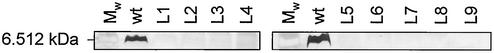
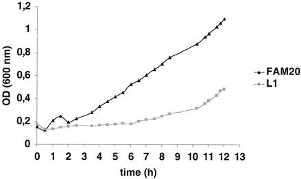
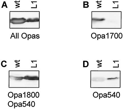
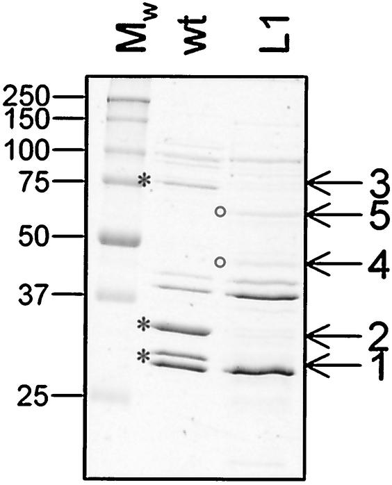
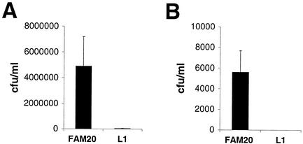
Molecular interaction between host mucosal surfaces and outer membrane components of microbes is crucial in the infection process. The outer membrane of pathogenic Neisseria contains surface molecules such as pili, PilC, and Opa and a monolayer of lipooligosaccharide (LOS), all of which are involved in the interaction with host cells. Pili mediate the initial attachment to human epithelial cells, which is followed by tight contact between bacteria and the eucaryotic cells, leading to bacterial invasion. To further examine the basis for bacterium-host cell contact, we constructed an LOS-deficient Neisseria meningitidis serogroup C mutant. LOS deficiency was without exception accompanied by altered colony opacity and morphology, which most likely represented an "on" switch for Opa540 expression, and by reduced levels of the iron-regulated proteins FetA and FbpA. We show here that LOS is essential for pilus-associated adherence but dispensable for fiber formation and twitching motility. The absence of attachment to epithelial cells could not be attributed to altered levels of piliation or defects in the pilus adhesion phenotype. Further, LOS mutants do not invade host cells and have lost the natural competence for genetic transformation.


Figures

FIG. 1.
FIG. 1.
SDS-PAGE analysis of LOS expressed by N. meningitidis FAM20 (wt) and its lpxA mutants (L1 to L9). Proteinase K-digested whole-cell lysates were separated on Tricine polyacrylamide gels and silver stained. Mw, molecular weight marker.
FIG. 2.
FIG. 2.
Growth curves of the wild-type FAM20 and the lpxA mutant L1. Bacteria were grown in GC medium with Kellogg's supplement. The same growth retardation was observed for mutants L2 to L9 (data not shown).
FIG. 3.
FIG. 3.
Western immunoblots of outer membranes of FAM20 (wt) and its lpxA mutant L1 incubated with anti-Opa monoclonal antibody (MAb) 4B12/C11 recognizing all Opas (A), anti-Opa MAb H21.1, recognizing Opa1700 (B), anti-Opa MAb H22.2, recognizing Opa1800 and Opa540 (C), and anti-Opa MAb 7-24-D9, recognizing Opa540 (D). Mutant L1 lost Opa1700 expression and gained Opa540 expression. A faint band reacting with Opa540 antibodies was often, but not always, observed in the wild type and could represent cross-reaction of the antibody with Opa1700 and Opa1800 or a minor population of wild-type bacteria that spontaneously turned on Opa540 by phase variation. The criteria for turn-on of Opa540 in L1 are the colony color difference between the wild type (weakly opaque) and L1 (strongly opaque and yellowish) and the very distinct Opa540 signal in the L1 mutant.
FIG. 4.
FIG. 4.
Analysis of outer membrane proteins expressed by N. meningitidis FAM20 (wt) and its lpxA mutant L1. Outer membrane proteins were purified by the LiAc method for outer membrane bleb purification and separated by SDS-PAGE. Five proteins with altered expression levels in the lpxA mutant were subjected to amino-terminal sequencing. Asterisks, proteins with reduced levels in the LOS mutants; circles, proteins with increased amounts in the mutants. Identical results were obtained with outer membranes prepared by sarcosyl extraction. The same profiles were observed with the other eight mutants (L2 to L9; data not shown). Mw, molecular weight marker.
FIG. 5.
FIG. 5.
LOS-deficient N. meningitidis fails to interact with human epithelial cells. (A) FAM20 and its lpxA-deficient mutant L1 were allowed to adhere to confluent layers of ME180 cells for 90 min. The cell layers were washed, treated with saponin, and spread on GC plates. (B) Invasion into ME180 cells of FAM20 and its lpxA-deficient mutant L1 was assessed for 6 h. Extracellular bacteria were killed by gentamicin, and the cell layers were treated with saponin and spread on GC plates. Mean values and standards deviations of three independent experiments are shown. Identical results were obtained with mutants L2 to L9 (data not shown).
FIG. 6.
FIG. 6.
(A) Transmission electron micrographs showing piliation of meningococcal strain FAM20 (wt) and its LOS-deficient mutant L1. (B) Immunoblotting of purified pili and outer membrane (OM) preparations by using PilC-specific antiserum. The same results were obtained with mutants L2 to L9 (data not shown).

References

    1. Billker, O., A. Popp, S. D. Gray-Owen, and T. F. Meyer. 2000. The structural basis of CEACAM-receptor targeting by neisserial Opa proteins. Trends Microbiol. 8:258-260. - PubMed
    1. Carson, S. D., P. E. Klebba, S. M. Newton, and P. F. Sparling. 1999. Ferric enterobactin binding and utilization by Neisseria gonorrhoeae. J. Bacteriol. 181:2895-2901. - PMC - PubMed
    1. Carson, S. D., B. Stone, M. Beucher, J. Fu, and P. F. Sparling. 2000. Phase variation of the gonococcal siderophore receptor FetA. Mol. Microbiol. 36:585-593. - PubMed
    1. Chen, I., and E. C. Gotschlich. 2001. ComE, a competence protein from Neisseria gonorrhoeae with DNA-binding activity. J. Bacteriol. 183:3160-3168. - PMC - PubMed
    1. Darzins, A. 1993. The pilG gene product, required for Pseudomonas aeruginosa pilus production and twitching motility, is homologous to the enteric single-domain response regulator CheY. J. Bacteriol. 175:5934-5944. - PMC - PubMed

Publication types

MeSH terms

LinkOut - more resources