

THP-1 cell apoptosis in response to Mycobacterial infection
- PMID: 12496173
- PMCID: PMC143334
- DOI: 10.1128/IAI.71.1.254-259.2003
THP-1 cell apoptosis in response to Mycobacterial infection
Abstract
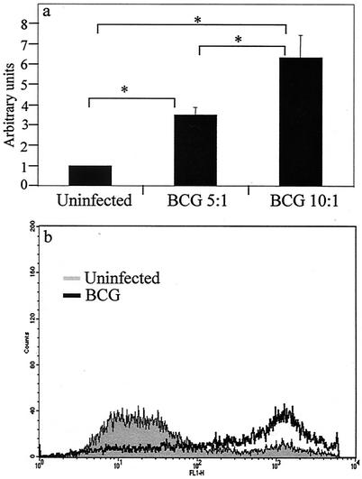
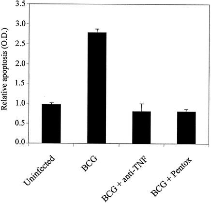
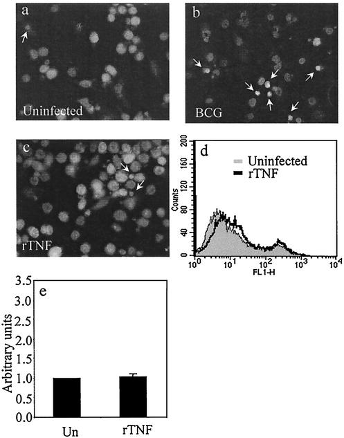
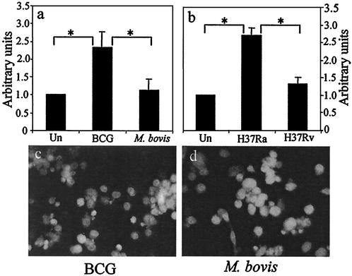
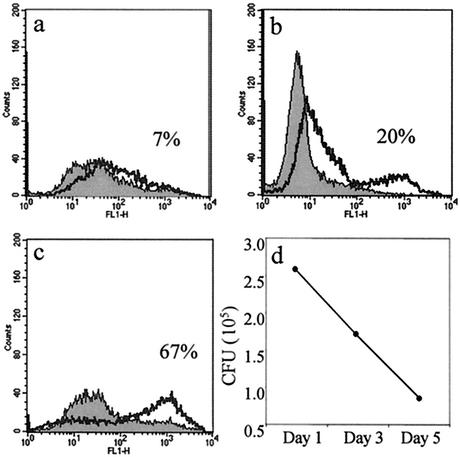
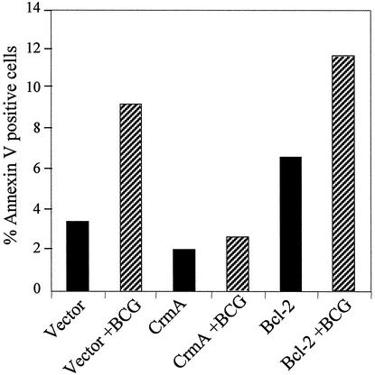
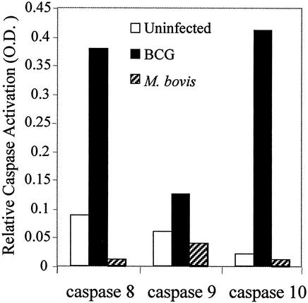
We previously reported that Mycobacterium tuberculosis infection primes human alveolar macrophages (HAM) for tumor necrosis factor alpha (TNF-alpha)-mediated apoptosis and that macrophage apoptosis is associated with killing internalized bacilli. Virulent mycobacterial strains elicit much less apoptosis than attenuated strains, implying that apoptosis is a defense against intracellular infection. The present study evaluated the potential for phorbol myristate acetate-differentiated THP-1 cells to mimic this response of primary macrophages. Consistent with the behavior of alveolar macrophages, attenuated M. tuberculosis H37Ra and Mycobacterium bovis BCG strongly induce THP-1 apoptosis, which requires endogenous TNF. THP-1 apoptosis is associated with reduced viability of infecting BCG. In contrast, virulent wild-type M. tuberculosis H37Rv and M. bovis do not increase THP-1 apoptosis over baseline. BCG induced early activation of caspase 10 and 9, followed by caspase 3. In contrast, wild-type M. bovis infection failed to activate any caspases in THP-1 cells. BCG-induced THP-1 apoptosis is blocked by retroviral transduction with vectors expressing crmA but not bcl-2. We conclude that differentiated THP-1 cells faithfully model the apoptosis response of HAM. Analysis of the THP-1 cell response to infection with virulent mycobacteria suggests that TNF death signals are blocked proximal to initiator caspase activation, at the level of TNF receptor 1 or its associated intracytoplasmic adaptor complex. Interference with TNF death signaling may be a virulence mechanism that allows M. tuberculosis to circumvent innate defenses leading to apoptosis of infected host cells.
Figures
References
-
- Balcewicz-Sablinska, M. K., J. Keane, H. Kornfeld, and H. G. Remold. 1998. Pathogenic Mycobacterium tuberculosis evades apoptosis of host macrophages by release of TNF-R2, resulting in inactivation of TNF-alpha. J. Immunol. 161:2636-2641. - PubMed
-
- Duan, L., H. Gan, J. Arm, and H. G. Remold. 2001. Cytosolic phospholipase A2 participates with TNF-alpha in the induction of apoptosis of human macrophages infected with Mycobacterium tuberculosis H37Ra. J. Immunol. 166:7469-7476. - PubMed
-
- Fratazzi, C., R. D. Arbeit, C. Carini, and H. G. Remold. 1997. Programmed cell death of Mycobacterium avium serovar 4-infected human macrophages prevents the mycobacteria from spreading and induces mycobacterial growth inhibition by freshly added, uninfected macrophages. J. Immunol. 158:4320-4327. - PubMed
-
- Gasperi, C., M. Rescigno, F. Granucci, S. Citterio, M. K. Matyszak, M. T. Sciurpi, L. Lanfrancone, and P. Ricciardi-Gastagnoli. 1999. Retroviral gene transfer, rapid selection, and maintenance of the immature phenotype in mouse dendritic cells. J. Leukoc. Biol. 66:263-267. - PubMed
Publication types
MeSH terms
Substances
Grants and funding
LinkOut - more resources
Full Text Sources
Other Literature Sources
Medical
Research Materials
