. 2003 Jan;71(1):260-6.
doi: 10.1128/IAI.71.1.260-266.2003.

Entry of the lymphogranuloma venereum strain of Chlamydia trachomatis into host cells involves cholesterol-rich membrane domains

Affiliations

Entry of the lymphogranuloma venereum strain of Chlamydia trachomatis into host cells involves cholesterol-rich membrane domains

Isabelle Jutras et al. Infect Immun. 2003 Jan.

Abstract

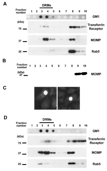
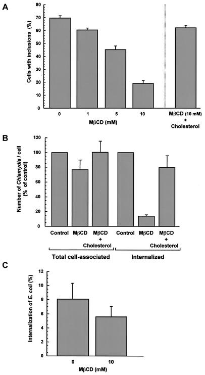
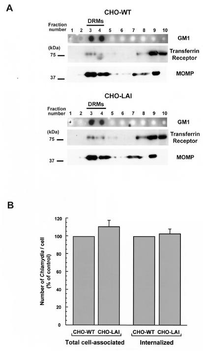
Chlamydiae are bacterial pathogens which develop strictly inside the epithelial cells of their hosts. The mechanism used by chlamydiae to enter cells is not well characterized; however, it is thought to consist of a receptor-mediated process. In addition, the formation of clathrin-coated pits appears to be dispensable for chlamydiae to be internalized by host cells. Clathrin-independent endocytosis has recently been shown to occur through cholesterol-rich lipid microdomains, which are characterized by detergent insolubility. In the present study, we investigated whether these lipid domains play a role in Chlamydia trachomatis serovar L2 internalization by host cells. Our results show that after binding to HeLa cells, chlamydiae are associated with detergent-resistant lipid microdomains (DRMs), which can be isolated by fractionation of infected HeLa cells and flotation on a sucrose gradient. After internalization by HeLa cells, chlamydiae were still found in DRMs. In addition, extraction of plasma membrane cholesterol inhibited infection of HeLa cells by C. trachomatis. Many of the proteins associated with DRMs are glycosylphosphatidylinositol (GPI)-anchored proteins; however, our results could not identify a role for GPI-anchored proteins in the entry process. The same results were obtained for Chlamydia psittaci strain GPIC. We propose that cholesterol-rich domains participate in the entry of chlamydiae into host cells. Chlamydia binding to cholesterol-rich domains may lead to coalescence of the bacterial cells, which could trigger internalization by host cells.


Figures

FIG. 1.
FIG. 1.
Chlamydiae associate with DRMs during infection of HeLa cells. (A) HeLa cells were incubated with C. trachomatis for 90 min at 4°C prior to cellular fractionation. Ganglioside GM1 staining, detected by dot blotting 2 μg of each fraction with HRP-conjugated cholera toxin, indicated the low-density fractions containing DRMs (fractions 3 to 5). Equivalent amounts of proteins from each fraction were resolved by SDS-PAGE. The same membrane was Western blotted with antibodies against the transferrin receptor or Rab5 (found in fractions 8 to 10, which contained the bulk of the solubilized membrane proteins) or against C. trachomatis MOMP. (B) A suspension of C. trachomatis alone was incubated in 1% Triton X-100 and fractionated on a sucrose gradient. The total content of each fraction was resolved by SDS-PAGE and Western blotted against MOMP. (C) DRMs prepared from Chlamydia-infected cells as described above for panel A were pooled, and the bacteria were pelleted. The recovered bacteria were washed in PBS and incubated with HeLa cells for 20 h at 37°C. Infected cells were fixed and immunofluorescently labeled with an anti-Chlamydia antibody to reveal the intracellular inclusions. (D) HeLa cells were incubated with C. trachomatis for 5 h at 37°C prior to fractionation. Ganglioside GM1, the transferrin receptor, Rab5, and MOMP were detected as described above for panel A.
FIG. 2.
FIG. 2.
Extraction of membrane cholesterol inhibits Chlamydia entry. (A) HeLa cells were incubated in the absence of MβCD or in the presence of different concentrations of MβCD for 30 min at 37°C. The cells either were immediately incubated with C. trachomatis at an MOI of 3 for 90 min at room temperature or were treated with 800 μg of cholesterol per ml for 12 min at 37°C prior to infection (right bar). The cells were washed to remove unbound bacteria and were incubated for an additional 16 h at 37°C. The cells were fixed, and Chlamydia inclusions were immunofluorescently labeled. (B) HeLa cells were incubated in the absence of MβCD or in the presence of 10 mM MβCD for 30 min at 37°C. MβCD was removed, and the cells were not treated or treated with 800 μg of cholesterol per ml for 12 min at 37°C. The cells were then infected with C. trachomatis at an MOI of 3 for 90 min at room temperature, washed, and incubated for an additional 3 h at 37°C. The cells were fixed and immunofluorescently labeled to visualize cell-associated and internalized bacteria. (C) HeLa cells were incubated in the absence of MβCD or in the presence of 10 mM MβCD for 30 min at 37°C and infected with E. coli MC4100/pRI203 for 2 h at room temperature. Unbound bacteria were removed, and the cells were incubated for an additional 90 min at 37°C. Extracellular bacteria were killed by incubating the cells in the presence of 50 μg of gentamicin per ml for 90 min at 37°C. The cells were rinsed with PBS and lysed in 0.1% Triton X-100. Released bacteria were titrated, and bacterial internalization was calculated by determining the percentage of viable counts.
FIG. 3.
FIG. 3.
(A) Chlamydiae associate with the DRMs of CHO cells deficient in GPI-anchored proteins. Wild-type CHO cells (CHO-WT) or GPI-anchored protein-deficient CHO cells (CHO-LAI) were incubated with C. trachomatis for 90 min at 4°C prior to isolation of DRMs. Ganglioside GM1 staining indicated the low-density fractions containing DRMs (fractions 3 and 4). Equivalent amounts of proteins from each fraction were resolved by SDS-PAGE. The same membrane was Western blotted with an antibody against the transferrin receptor or against C. trachomatis MOMP. (B) Chlamydiae efficiently enter CHO cells deficient in GPI-anchored proteins. Wild-type CHO or CHO-LAI cells were infected with C. trachomatis for 90 min at 37°C. Unbound chlamydiae were removed, and the cells were incubated for an additional 6 h at 37°C. The cells were fixed, and the chlamydiae were immunofluorescently labeled to visualize cell-associated or internalized bacteria.

References

    1. Abrami, L., M. Fivaz, T. Kobayashi, T. Kinoshita, R. G. Parton, and F. G. van der Goot. 2001. Cross-talk between caveolae and glycosylphosphatidylinositol-rich domains. J. Biol. Chem. 276:30729-30736. - PubMed
    1. Anderson, H. A., Y. Chen, and L. C. Norkin. 1996. Bound simian virus 40 translocates to caveolin-enriched membrane domains, and its entry is inhibited by drugs that selectively disrupt caveolae. Mol. Biol. Cell 7:1825-1834. - PMC - PubMed
    1. Baorto, D. M., Z. Gao, R. Malaviya, M. L. Dustin, A. van der Merwe, D. M. Lublin, and S. N. Abraham. 1997. Survival of FimH-expressing enterobacteria in macrophages relies on glycolipid traffic. Nature 389:636-639. - PubMed
    1. Boleti, H., A. Benmerah, D. M. Ojcius, N. Cerf-Bensussan, and A. Dautry-Varsat. 1999. Chlamydia infection of epithelial cells expressing dynamin and Eps15 mutants: clathrin-independent entry into cells and dynamin-dependent productive growth. J. Cell Sci. 112:1487-1496. - PubMed
    1. Brown, D. A., and E. London. 2000. Structure and function of sphingolipid- and cholesterol-rich membrane rafts. J. Biol. Chem. 275:17221-17224. - PubMed

Publication types