. 2003 Jan;71(1):411-7.
doi: 10.1128/IAI.71.1.411-417.2003.

Roles of the Maltese cross form in the development of parasitemia and protection against Babesia microti infection in mice

Affiliations

Roles of the Maltese cross form in the development of parasitemia and protection against Babesia microti infection in mice

Naoaki Yokoyama et al. Infect Immun. 2003 Jan.

Abstract

Babesia microti, a hemoprotozoan parasite of rodents, is also important as a zoonotic agent of human babesiosis. The Maltese cross form, which consists of four masses in an erythrocyte, is characteristic of the developmental stage of B. microti. Monoclonal antibody (MAb) 2-1E, which specifically recognizes the Maltese cross form of B. microti, has been described previously. In the present study, we examined the roles of the Maltese cross form during the infectious course of B. microti in mice. The number of the Maltese cross form increased in the peripheral blood of infected mice prior to the peak of parasitemia. With confocal laser scanning microscopy, MAb 2-1E was found to be reactive with the ring form, with the parasites undergoing transformation to the Maltese cross form and subsequent division, and also with extracellular merozoites. Furthermore, the Maltese cross form-related antigen (MRA) gene was isolated from a B. microti cDNA library by immunoscreening with MAb 2-1E, and the nucleotide sequence was determined. Genomic analyses indicated that the MRA gene exists as a single-copy gene in B. microti. Immunization of mice with recombinant MRA induced significant protective immunity against B. microti infection. These findings indicate that the Maltese cross form plays important roles in both the development of parasitemia and the protective response against the infection.


Figures
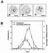
FIG. 1.
FIG. 1.
(A) Micrographs of intraerythrocytic Babesia merozoites in Giemsa-stained blood smears. The ring (panel a), pear-shaped (panel b), and Maltese cross (panel c) forms were observed in the B. microti-infected murine erythrocytes. Bars = 2 μm. (B) Appearance curve for the B. microti Maltese cross (MC) form. Three female BALB/c mice that were approximately 8 weeks old were each infected intraperitoneally with 1 × 10⁷ B. microti-infected erythrocytes. The parasitemia and the percentage of the Maltese cross form in infected erythrocytes were monitored daily by examining blood smears periodically prepared from blood from the tail veins. Each symbol indicates the mean, and the error bars indicate the standard errors.
FIG. 2.
FIG. 2.
IFAT performed by using confocal laser scanning microscopy. Methanol-fixed smears of the B. microti-infected erythrocytes were incubated with MAb 2-1E. The MAb-antigen reaction (green) was visualized with the FITC-conjugated secondary antibody. Bars = 5 μm.
FIG. 3.
FIG. 3.
(A) Deduced amino acid sequence encoded by the MRA gene (GenBank accession number AB079857). The sequence region showing significant identity to the sequence encoded by the BMN 1-15 gene is indicated by underlining. (B) Alignment of the deduced amino acid sequence encoded by the MRA gene with the amino acid sequence of BMN 1-15 of B. microti (GenBank accession number AF206525). Amino acid gaps are indicated by dashes, amino acid identities are indicated by colons, and conservative changes are indicated by dots.
FIG. 4.
FIG. 4.
(A) Southern blot analyses. The B. microti genomic DNA was digested with HindIII (lane 1), PstI (lane 2), XbaI (lane 3), or EcoRI (lane 4), and the separated DNA fragments were hybridized with the MRA gene probe. The positions of λ HindIII DNA size markers (in kilobase pairs) are indicated on the left. (B) PCR amplification of the MRA gene in the DNA extracted from normal blood (lane 1) or B. microti-infected blood (lane 2) and cDNA clone of the MRA gene (lane 3). Lane M contained λ HindIII DNA size markers.
FIG. 5.
FIG. 5.
(A) IFAT. Methanol-fixed smears of AcMRA-infected (panel a) or AcFB-D-infected (panel b) Sf9 cells were incubated with immune serum collected from a B. microti-infected mouse. The MAb-antigen reaction (white) was visualized with the FITC-conjugated secondary antibody. Bars = 20 μm. (B) Western blot analyses. The antigens prepared from noninfected (lane 1), AcMRA-infected (lane 2), or AcFB-D-infected (lane 3) Sf9 cell lysate or purified B. microti lysate (lane 4) were reacted with MAb 2-1E and visualized with an ECL kit. The positions of the molecular mass standards (in kilodaltons) are indicated on the left.
FIG. 6.
FIG. 6.
IFAT performed by using confocal laser scanning microscopy. Methanol-fixed smears of B. microti-infected erythrocytes were incubated with immune sera of the MRA (a) or FB-D (b) group. The MAb-antigen reaction (green) was visualized with the FITC-conjugated secondary antibody. Bar = 5 μm (a) or 10 μm (b).
FIG. 7.
FIG. 7.
Vaccine efficacy of the recombinant MRA against B. microti challenge. The five mice in the MRA group and the five mice in the FB-D group were immunized three times with AcMRA-infected cell lysate and AcFB-D-infected cell lysate, respectively, while the control group did not receive any immunogen. After challenge with 1 × 10⁷ B. microti-infected erythrocytes, the developmental parasitemia in each group was monitored daily by examining blood smears periodically prepared from blood from the tail veins. Each symbol indicates the mean, and the error bars indicate the standard errors. The asterisks indicate the days on which there was a significant difference (P < 0.05) between the MRA and FB-D groups. The data are representative of two separate experiments.

Comment in

Similar articles

Cited by

References

    1. Avarzed, A., I. Igarashi, D. T. De Waal, S. Kawai, Y. Oomori, N. Inoue, Y. Maki, Y. Omata, A. Saito, H. Nagasawa, Y. Toyoda, and N. Suzuki. 1998. Monoclonal antibody against Babesia equi: characterization and potential application of antigen for serodiagnosis. J. Clin. Microbiol. 36:1835-1839. - PMC - PubMed
    1. Benach, J. L., and G. S. Habicht. 1981. Clinical characteristics of human babesiosis. J. Infect. Dis. 144:481. - PubMed
    1. Daly, T. M., and C. A. Long. 1996. Influence of adjuvants on protection induced by a recombinant fusion protein against malarial infection. Infect. Immun. 64:2602-2608. - PMC - PubMed
    1. Eskow, E. S., P. J. Krause, A. Spielman, K. Freeman, and J. Aslanzadeh. 1999. Southern extension of the range of human babesiosis in the eastern United States. J. Clin. Microbiol. 37:2051-2052. - PMC - PubMed
    1. Fawcett, D. W., P. A. Conrad, J. G. Grootenhuis, and S. P. Morzaria. 1987. Ultrastructure of the intraerythrocytic stage of Theileria species from cattle and waterbuck. Tissue Cell 19:643-655. - PubMed

Publication types

MeSH terms

Associated data

LinkOut - more resources