. 2003 Jan;71(1):516-23.
doi: 10.1128/IAI.71.1.516-523.2003.

Role of sarA in the pathogenesis of Staphylococcus aureus musculoskeletal infection

Affiliations

Role of sarA in the pathogenesis of Staphylococcus aureus musculoskeletal infection

Jon S Blevins et al. Infect Immun. 2003 Jan.

Abstract

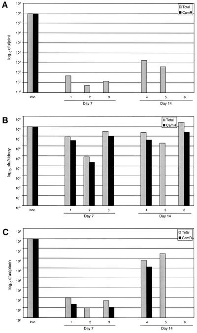
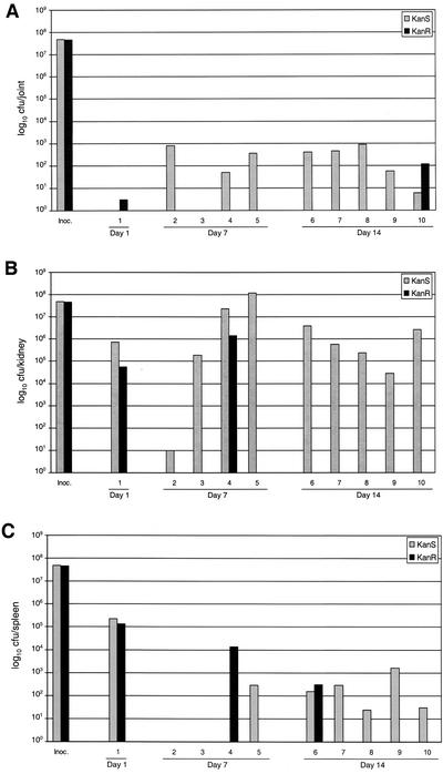
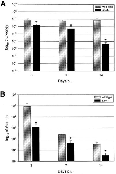
We recently demonstrated that mutation of sarA in clinical isolates of Staphylococcus aureus results in a phenotype that is distinct by comparison to sarA mutants generated in the laboratory strain RN6390 (J. S. Blevins, K. E. Beenken, M. O. Elasri, B. K. Hurlburt, and M. S. Smeltzer, Infect. Immun. 70:470-480, 2002). This raises the possibility that studies demonstrating that RN6390 sarA mutants are attenuated do not accurately reflect the role of sarA in the pathogenesis of staphylococcal disease. To test this hypothesis, we used a murine model of musculoskeletal infection to assess the virulence of sarA and agr mutants generated in a clinical isolate of S. aureus (UAMS-1). By using this model, we confirmed that mutation of sarA and/or agr results in a reduced capacity to cause both septic arthritis and osteomyelitis.


Figures

FIG. 1.
FIG. 1.
Histological evidence of musculoskeletal infection. Photomicrographs of H&E-stained sections 14 days after infection with S. aureus. (A) Section scored as unaffected; inflammation score = 0. (B) Section showing a joint with mild synovitis; 1+ inflammation with a small joint abscess (AJ) and no demonstrable bone involvement. (C) Section with moderate synovitis and mild osteomyelitis; 2+ inflammation in joint and 1+ inflammation in bone. The severity of synovial inflammation has led to erosion of articular cartilage (EAC) and cortical bone (ECB) and secondary osteomyelitis with physis destruction (PD). (D) Section showing a hind limb with severe synovitis and osteomyelitis; 3+ inflammation scores in both the bone and synovium. In addition to the aforementioned parameters, two bone abscesses (AB) are also present, one in the tibia (upper bone) and the other in the femur (lower bone). The inset shows a Gram-stained serial section with gram-positive cocci present in the subchondral bone of the tibia. Magnifications: panels A to D, ×16; panel D inset, ×200.
FIG. 2.
FIG. 2.
Histological evidence of primary osteomyelitis. Photomicrographs of H&E- and Gram-stained sections 14 days after infection with S. aureus. (A) Section scored as negative for synovitis with 3+ inflammation in the bone, abscess formation (AB), and physis destruction (PD). To maintain orientation, the AB and PD labels are in the same relative positions in each panel. Magnification, ×20. (B) Higher magnification, ×200, of the section shown in panel A. (C) Gram staining of a serial section demonstrating the presence of bacteria in the abscess; magnification, ×200. The framed region in panels B and C coincides with the inset; magnification ×400.
FIG. 3.
FIG. 3.
Plasmid stability in vivo. Mice were infected with the pSARA-complemented UAMS-1 sarA mutant (UAMS-969). Samples were obtained from a joint (A), a kidney (B), and the spleen (C) at days 7 and 14 p.i. Results are reported as the total number of CFU recovered and the number that were chloramphenicol resistant (Camr). Inoc., inoculum.
FIG. 4.
FIG. 4.
Coinfection studies. Mice were coinfected with equal numbers of cells of UAMS-1 and its isogenic sarA mutant (UAMS-929). One mouse died on day 1 p.i. All other samples from a joint (A), a kidney (B), and the spleen (C) were taken at 7 and 14 days p.i. Results are reported as the number of colonies that were sensitive to kanamycin-neomycin (Kans) and the number that were resistant (Kanr). Inoc., inoculum.
FIG. 5.
FIG. 5.
Impact of sarA mutation on colonization of and survival in mouse tissues. Mice infected intravenously with UAMS-1 or its isogenic sarA mutant (UAMS-929) were euthanized at days 3, 7, and 14, and bacteria were isolated from a kidney (A) and the spleen (B). Results are reported as the mean number of CFU ± the standard error of the mean, and each mean represents a group of five mice. The asterisk indicates a statistically significant difference (P < 0.05).

References

    1. Abdelnour, A., S. Arvidson, T. Bremell, C. Ryden, and A. Tarkowski. 1993. The accessory gene regulator (agr) controls Staphylococcus aureus virulence in a murine arthritis model. Infect. Immun. 61:3879-3885. - PMC - PubMed
    1. Bischoff, M., J. M. Entenza, and P. Giachino. 2001. Influence of a functional sigB operon on the global regulators sar and agr in Staphylococcus aureus. J. Bacteriol. 183:5171-5179. - PMC - PubMed
    1. Blevins, J. S., K. E. Beenken, M. O. Elasri, B. K. Hurlburt, and M. S. Smeltzer. 2001. Strain-dependent differences in the regulatory roles of sarA and agr in Staphylococcus aureus. Infect. Immun. 70:470-480. - PMC - PubMed
    1. Blevins, J. S., A. F. Gillaspy, T. M. Rechtin, B. K. Hurlburt, and M. S. Smeltzer. 1999. The staphylococcal accessory regulator (sar) represses transcription of the Staphylococcus aureus collagen adhesin gene (cna) in an agr-independent manner. Mol. Microbiol. 33:317-326. - PubMed
    1. Booth, M. C., R. V. Atkuri, S. K. Nanda, J. J. Iandolo, and M. S. Gilmore. 1995. Accessory gene regulator controls Staphylococcus aureus virulence in endophthalmitis. Investig. Ophthalmol. Vis. Sci. 36:1828-1836. - PubMed

Publication types

MeSH terms