. 2002 Dec 23;159(6):983-91.
doi: 10.1083/jcb.200208172. Epub 2002 Dec 23.

The prepower stroke conformation of myosin V

Affiliations

The prepower stroke conformation of myosin V

Stan Burgess et al. J Cell Biol. .

Abstract

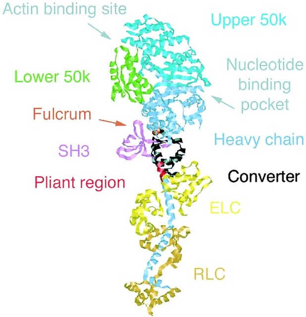
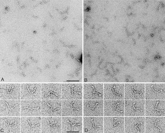
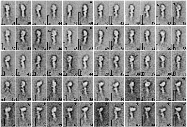
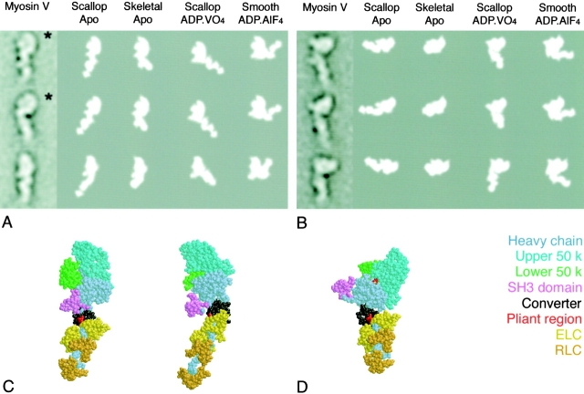
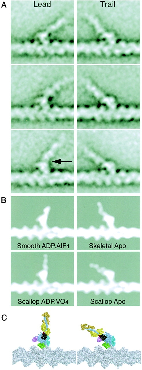
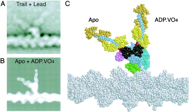
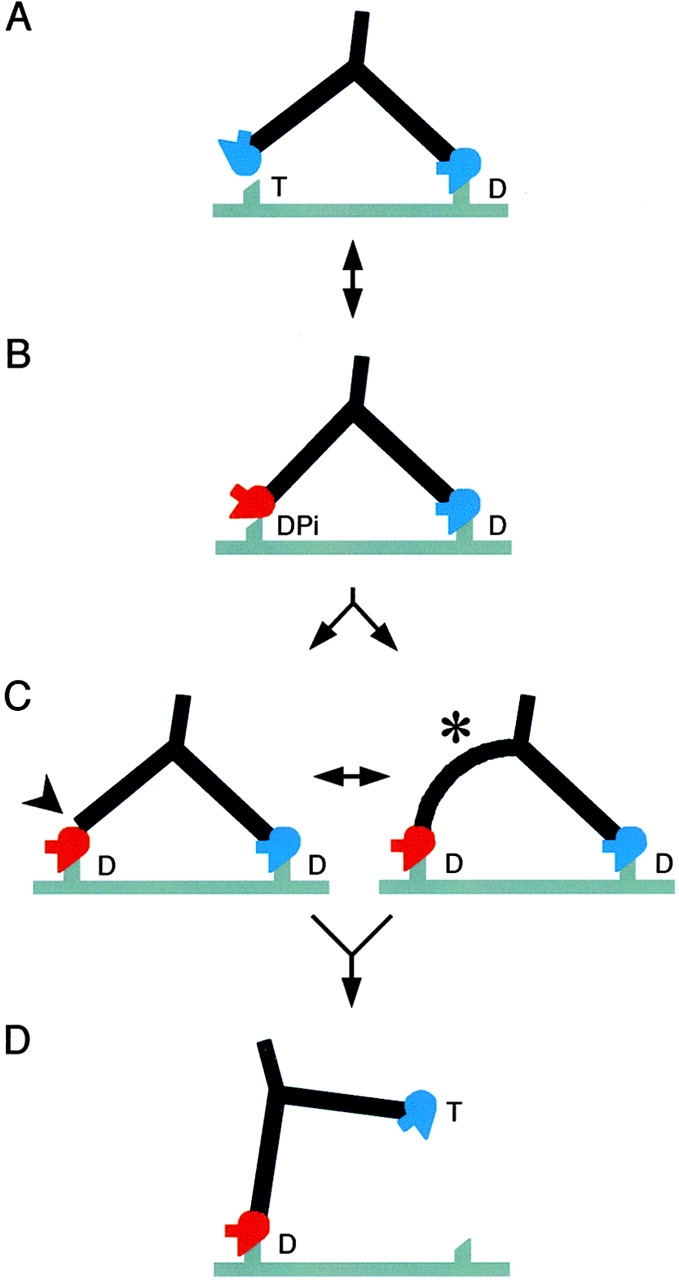
We have used electron microscopy and single-particle image processing to study head conformation in myosin V molecules. We find that in the presence of ATP, many heads have a sharply angled conformation that is rare in its absence. The sharply angled conformation is similar to a myosin II atomic structure proposed to mimic the prepower stroke state. The leading head in molecules attached to actin by both heads has a similar conformation, but is also sharply angled in a second plane by tethering through the trail head. The lead head lever joins the motor domain approximately 5 nm axially from where it joins the trail motor. These positions locate the converter subdomain and show the lead motor is in the prepower stroke conformation. Tethering by the trail head places the lead head motor domain at the correct axial position along the actin for binding, but at the wrong orientation. Attachment is achieved either by bending the lead head lever throughout its length or at the pliant point. The microscopy shows that most of the walking stride is produced by changes in lever angle brought about by converter movement, but is augmented by distortion produced by thermal energy.


Figures

Figure 1.
Figure 1.
Crystal structure of the scallop myosin II apoenzyme. The motor domain is formed by the heavy chain and contains the nucleotide binding pocket and actin binding site. A cleft divides upper and lower 50-kD subdomains, and the SH3 β-barrel of the NH2-terminal subdomain is prominent. The COOH-terminal element of the converter is an α-helix extending into the lever, which is complexed by the essential and regulatory light chains (ELC and RLC, respectively). Between the converter and ELC, a few residues of the heavy chain form a pliant region allowing the lever to bend relative to the motor. The fulcrum for ATP-driven lever movement is within the motor domain (orange spacefilled residue). This and all subsequent atomic diagrams were made using RasMol (Sayle and Milner-White, 1995).
Figure 2.
Figure 2.
Negatively stained myosin V HMM molecules. Fields of molecules in the absence (A) and presence (B) of nucleotide (100 μM ATP; 20°C). Galleries of molecules in the absence (C) and presence (D) of nucleotide. We consistently found image quality to be better without nucleotide. Bars: 100 nm (A and B); 50 nm (C and D).
Figure 3.
Figure 3.
Image averages of myosin V heads in the absence and presence of ATP. Aligned images from both conditions (n = 1,172 in each case) were pooled and classified into 60 classes. Thus, each class has both apo and nucleotide images (where both exist). (top left to bottom right) Classes are presented in order of increasing value of n nucleotide /(n nucleotide+ n apo). At bottom right in each panel is the total number of images in the class. The histograms show the numbers of apo (left) or nucleotide (right) images in each class. The length of the lever is about half that observed in individual images. The remainder is lost due to variability in position of the COOH-terminal end. Classes in which n nucleotide/n apo >10 are marked with an asterisk. Motor domain morphology in these classes is occasionally seen in classes dominated by apo heads (black spots). Arrowheads indicate the cleft between lower and upper 50-kD subdomains, and the arrow (row 3) points to the nucleotide binding pocket. Panels are 25-nm wide.
Figure 4.
Figure 4.
Comparisons of myosin V heads with myosin II crystal structures. The most populous apo and nucleotide classes were compared with apo (skeletal and scallop) and transition state (scallop–ADP.VO4 and smooth–ADP.AlF4) crystal structures. Only features within motor domains were used for matching (see Results). In each row of A and B, the levers and converter subdomains of the four crystal structures are different, but the rest of the motor domains are very similar and resemble image average motor domains. (A) Apo myosin V motor domains show broadly two appearances (upper 2 and bottom classes) corresponding to two distinct motor orientations (shown in spacefill diagrams of the scallop apo structure in C). Asterisks mark evidence of flexibility at the motor domain–lever junction (see Results). (B) Nucleotide motor domains show broadly one type of appearance, which most closely resembles the scallop–ADP.VO4 structure (D).
Figure 5.
Figure 5.
Comparisons of myosin heads bound to actin with crystal structures of myosin II docked onto actin. (A) Image averages produced by classifying into six and four classes, respectively, 150 lead and 176 trail heads from molecules attached by both heads. Walking is toward the left in this and the following figures. (B) Models produced by docking head crystal structures with the atomic model of the actin filament (Lorenz et al., 1993) followed by low-pass filtering to 2-nm resolution. Note the similarity in appearance of the actin and myosin with A. (C) Colored models of scallop structures docked onto actin. The arrow marks the pliant point. The angle between the lever arm and the actin filament was 129–143° in lead heads and 39–49° in trail heads. Note also that the sharp bend of the scallop–ADP.VO4 structure is perpendicular to this view and does not show. The converter (black) and lever move in the plane of the page relative to the rest of the motor.
Figure 6.
Figure 6.
Demonstration that lead and trail heads have their converters in different positions. Composite images were produced by superposition of image averages (A) and atomic models of docked scallop apo and ADP.VO4 structures (B and C). Note the points of emergence of the levers (i.e., the pliant regions) differ by ∼5 nm in the image averages (compare with the actin subunit spacing) and 3.6 nm in the atomic models.
Figure 7.
Figure 7.
Structural cycle of myosin V walking. Red motor domains represent the converter and lever in the prepower stroke conformation, and blue ones represent the postpower stroke conformation. The actin filament is gray and shows two preferred attachment sites for heads spaced by 36 nm. Myosin (drawn to a similar scale) is walking left and has a head orientation that shows a prominent SH3 domain. T, DPi, and D (ATP, ADP + phosphate, and ADP) indicate the likely head nucleotide contents in vivo. Note that if ATP is limiting in vitro, as in our data with actin, then the head contents will be different (see Discussion). (A) The attached head is close to the rigor orientation; the motor domain of the second head can reach the next binding site on actin, but the apo-like head shape is wrong for attachment. (B) The detached head has switched to the angled conformation seen in unattached molecules (Fig. 4 B), which in this view appears straight (Fig. 5 B, scallop–ADP.VO4). The motor domain may attach weakly to actin, but is not oriented correctly for strong binding because the lever is not perpendicular to the actin filament. (C) Stereospecific attachment of the lead head motor to actin. We propose that attachment is achieved by distortion either at the lead head pliant region (arrowhead), or throughout its lever (asterisk). This explains why both straight and curved levers can be seen in lead head images, even though the converters are in the prepower stroke position. Note that little forward movement of the myosin tail accompanies attachment of the lead head. For simplicity, the subsequent actin-activated release of Pi is not shown as a separate step. Note also that though both heads contain only ADP, they have very different conformations. (D) Release of the trail head on binding ATP. The intramolecular strain is relieved, the lead lever becomes nearly perpendicular to the filament and the myosin tail moves forward. This would quickly be followed by the working stroke of the lead head, completing the cycle.

Similar articles

Cited by

References

    1. Bauer, C.B., H.M. Holden, J.B. Thoden, R. Smith, and I. Rayment. 2000. X-ray structures of the apo and MgATP-bound states of Dictyostelium discoideum myosin motor domain. J. Biol. Chem. 275:38494–38499. - PubMed
    1. Berg, J.S., B.C. Powell, and R.E. Cheney. 2001. A millennial myosin census. Mol. Biol. Cell. 12:780–794. - PMC - PubMed
    1. Burgess, S.A., M. Walker, H.D. White, and J. Trinick. 1997. Flexibility within myosin heads revealed by negative stain and single-particle analysis. J. Cell Biol. 139:675–681. - PMC - PubMed
    1. de la Cruz, E.M., A.L. Wells, S.S. Rosenfeld, E.M. Ostap, and H.L. Sweeney. 1999. The kinetic mechanism of myosin V. Proc. Natl. Acad. Sci. USA. 96:13726–13731. - PMC - PubMed
    1. de la Cruz, E.M., E.M. Ostap, and H.L. Sweeney. 2001. Kinetic mechanism and regulation of myosin VI. J. Biol. Chem. 276:32373–32381. - PubMed

Publication types