. 2002 Dec 20;13(18):2411-5.
doi: 10.1097/00001756-200212200-00007.

Neurite branching on deformable substrates

Affiliations

Neurite branching on deformable substrates

Lisa A Flanagan et al. Neuroreport. .

Abstract

The mechanical properties of substrates underlying cells can have profound effects on cell structure and function. To examine the effect of substrate deformability on neuronal cell growth, protein-laminated polyacrylamide gels were prepared with differing amounts of bisacrylamide to generate substrates of varying deformability with elastic moduli ranging from 500 to 5500 dyne/cm. Mouse spinal cord primary neuronal cells were plated on the gels and allowed to grow and extend neurites for several weeks in culture. While neurons grew well on the gels, glia, which are normally co-cultured with the neurons, did not survive on these deformable substrates even though the chemical environment was permissive for their growth. Substrate flexibility also had a significant effect on neurite branching. Neurons grown on softer substrates formed more than three times as many branches as those grown on stiffer gels. These results show that mechanical properties of the substrate specifically direct the formation of neurite branches, which are critical for appropriate synaptic connections during development and regeneration.


Figures

Fig. 1
Fig. 1
Characterization of polyacrylamide gel substrates and neuronal cultures. (a) Shear moduli (G′) of polyacrylamide gels are plotted as a function of increasing bisacrylamide cross-linker concentrations. (b) Schematic diagram depicting polyacrylamide gel substrates. The top view highlights that gels of differing deformability do not differ in polymer mass but only in number of cross-links. The relative differences between the size of a neuron and its processes and the pore size of the gels and thickness of the gel substrates are also shown (not drawn to scale). (c) Samples of matrigel (three different concentrations) or gels coated with matrigel (four different bisacrylamide cross-linker concentrations) were analyzed by Western blot with an antibody to laminin, a major component of matrigel. Gels of varying deformability do not differ in amount of matrigel coating. (d) Mouse primary spinal cord cultures on glass grow as dense cultures with a variety of neuronal subtypes (arrow and arrowhead) and underlying, very flat glial cells (double arrowhead). Scale bar = 50 μm.
Fig. 2
Fig. 2
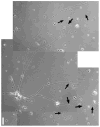
Spinal cord neurons on soft substrates. Neurons growing on a soft (0.08% bisacrylamide, 2300 dyne/cm2) gel extend neurites with multiple branch points (representative branches marked by arrows). Scale bar = 50 μm.
Fig. 3
Fig. 3
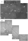
Spinal cord neurons on stiff substrates and glass. Neurons growing on (a) a stiff (0.2% bisacrylamide, 5500 dyne/cm2) gel and (b) glass as sparse cultures. Neurites are highlighted in black and representative branch points are marked by arrows. Scale bar = 50 μm.
Fig. 4
Fig. 4
Quantification of neurite branching on deformable substrates and glass. Neurite branches per mm of neurite are plotted as a function of increasing substrate stiffness. Glass is considered to have an infinite stiffness compared to the gels. Numbers next to points indicate the percentage of bisacrylamide used for gels of that stiffness. Numbers of branches were counted for all the neurons on gels from several different experiments (0.04% n = 3, 0.08% n = 4, 0.1% n = 3, 0.2% n = 3). Error bars are s.e.m.

References

    1. Hay ED. Am J Anat. 1982;165:1–12. - PubMed
    1. Bard JB, Hay ED. J Cell Biol. 1975;67:400–418. - PMC - PubMed
    1. Halliday NL, Tomasek JJ. Exp Cell Res. 1995;217:109–117. - PubMed
    1. Arora PD, Narani N, McCulloch CA. Am J Pathol. 1999;154:871–882. - PMC - PubMed
    1. Pelham RJ, Jr, Wang Y. Proc Natl Acad Sci USA. 1997;94:13661–13665. - PMC - PubMed

Publication types