. 2002 Dec 15;16(24):3136-41.
doi: 10.1101/gad.251202.

LIN-39/Hox triggers cell division and represses EFF-1/fusogen-dependent vulval cell fusion

Affiliations

LIN-39/Hox triggers cell division and represses EFF-1/fusogen-dependent vulval cell fusion

Gidi Shemer et al. Genes Dev. .

Abstract

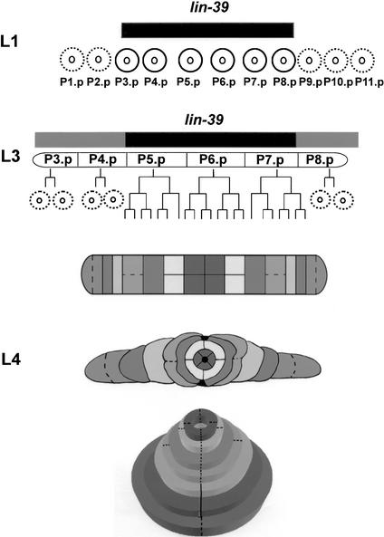
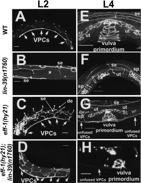
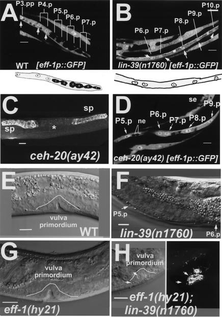
General mechanisms by which Hox genes establish cell fates are known. However, a few Hox effectors mediating cell behaviors have been identified. Here we found the first effector of LIN-39/HoxD4/Dfd in Caenorhabditis elegans. In specific vulval precursor cells (VPCs), LIN-39 represses early and late expression of EFF-1, a membrane protein essential for cell fusion. Repression of eff-1 is also achieved by the activity of CEH-20/Exd/Pbx, a known cofactor of Hox proteins. Unfused VPCs in lin-39(-);eff-1(-) double mutants fail to divide but migrate, executing vulval fates. Thus, lin-39 is essential for inhibition of EFF-1-dependent cell fusion and stimulation of cell proliferation during vulva formation. Supplemental material is available at http://www.genesdev.org.


Figures

Figure 1
Figure 1
lin-39 inhibits cell fusion during vulva formation. lin-39 is expressed and active at the first larval stage (L1) in six epidermal cells (P3.p-P8.p; black circles), also known as the vulva precursor cells (VPCs). This activity (black box above cells) is necessary and sufficient to inhibit the VPCs from adopting the fate of their neighboring cells [P(1,2,9–11).p]—fusion to the surrounding syncytium hyp7 (dashed circles). At L3, the VPCs are subjected to various signaling pathways. P(3,4,8).p undergo one cycle of division, followed by fusion of their daughter cells to hyp7 (dashed circles). In about half of the cases, P3.p does not divide before cell fusion. P(5–7).p escape cell fusion and continue to divide, yielding a 22-cell vulval primordium. It was proposed that basal activity of LIN-39 in P(3,4,8).p is not sufficient for these cells to escape cell fusion, and only elevated activity of LIN-39 induced by the Ras pathway in P(5–7).p in wild-type (Maloof and Kenyon 1998) or by ectopic Wnt signaling in all VPCs in some mutants (Gleason et al. 2002), allows these cells to escape cell fusion (black and shaded boxes above cells). Migration, cell fusion (dashed lines), ring formation, and invagination of the primordial cells at L4 lead to a tube-shaped adult vulva (Sharma-Kishore et al. 1999).
Figure 2
Figure 2
eff-1 is epistatic to lin-39 during early and late cell-fusion events. Confocal reconstructions of worms carrying an ajm-1∷GFP construct marking the adherens junctions of epithelial cells (Shemer et al. 2000; Koeppen et al. 2001). The worms were assayed at early L2 (AD) after the early fusion events of mid-L1 and at mid-L4 (EH) after the late fusion events of mid-L3 (see Fig. 1 for details). (A) Wild-type worms showing the six VPCs that escape fusion to the hypodermis after they attach to each other (n > 100). (E) Three of these VPCs escape cell fusion to hyp7 at L3, and their 22 great-granddaughters invaginate, forming a stack of seven rings (n > 100). (B,F) In lin-39(n1760) single null mutants, no VPCs formed after all of the Pn.p cells had fused to the hypodermis (*, n = 50), resulting in the absence of a vulva (Vul phenotype) at late L4 (F; n = 100). (C) eff-1(hy21) mutants grown at the restrictive temperature of 25°C showing the VPCs that fail to fuse with hyp7 (n = 50). P3.p, P4.p, and P8.p also fail to fuse in the L3 and attach to the vulva primordium formed from the descendants of P(5–7).p. (G) The result is a stack of rings connected to a row of ectopic cells (n = 70). Due to fusion failure in eff-1(hy21), ectopic dorsal epithelia (de) and lateral hypodermal seam cells (se) are present and migrate throughout the body of the worm. (D,H) Pn.p cells fail to fuse in eff-1(hy21);lin-39(n1760) double mutants at L1 (D; n = 45) and later at L3 (H; n = 28). The unfused VPCs migrate and invaginate, forming a “pseudo” vulval primordium that is incomplete and abnormal structurally, resulting in a nonfunctional vulva (cf. H and G,E). ut, uterus; sp, spermatheca. Anterior is to the left and dorsal is up, except for B, a ventral view. The fusion status of the cells was also confirmed by staining worms with the MH27 antibody (Podbilewicz and White 1994). Arrows mark unfused cells. Bar, 10μm.
Figure 3
Figure 3
lin-39 and ceh-20 repress eff-1 expression. (A,B) Z-serial sections from confocal reconstructions of L3 worms showing expression of eff-1promoter∷GFP (white) in vulval primordial cells in the presence (A) and absence (B) of lin-39 activity. (A) White arrows mark nuclei of cells that fuse with the surrounding hypodermal syncytium hyp7. All of these nuclei express eff-1 (arrows and white circles in the diagram beneath; nucleoli are small circles within nuclei, n = 35). Daughter cells of P(5–7).p, which escape fusion to the hypodermis due to lin-39 activity, do not express eff-1. (B) In the absence of lin-39 activity, all of the VPCs express eff-1 [P(6–10).p in this picture; see arrows and white nuclei in the diagram beneath; n = 40]. (C,D) ceh-20 inhibits cell fusion by repressing eff-1 expression. (C) ceh-20 L4 mutant stained with MH27 showing that in the absence of ceh-20, no vulva (*) is formed as a result of VPC fusion (n = 18). The uterine cells are shown in the weak staining between the spermathecae (sp). (D) Expression of eff-1promoter∷GFP (white) in vulval primordial cells in the absence of ceh-20 activity. These cells express GFP [P(5–9).p in this picture]. Seam cells (se) and ventral neural cells (ne) are also shown (n = 22). (EH) Nomarski micrographs showing vulval nuclei during organogenesis. (E) Wild-type worm at late L4. This vulva primordium is comprised of 22 nuclei (white line, invagination with 10 nuclei in this focal plane; n > 100). (F) lin-39(n1760) null mutant worm at early L4 showing only P5.p and P6.p that in the absence of lin-39 activity fused to hyp7 (n = 30). (G) eff-1(hy21) worm at exactly the same stage as that in E (white line, invagination; n = 60). (H) In the absence of lin-39, the VPCs do not proliferate in eff-1(hy21);lin-39(n1760) mutants (n = 34). However, the cells that fail to fuse in the absence of eff-1 activity, migrate and ultimately invaginate (white line). Right image shows a stack of four vulval rings (ajm-1∷GFP) from the same vulva. Anterior is left and dorsal is up, except for A, C, and D, which are ventral views. All worms were grown at 25°C for complete penetrance of the eff-1(hy21) mutation. Bar, 10μm.
Figure 4
Figure 4
A model of lin-39 activity during vulva formation. (A) lin-39 inhibits cell fusion in the VPCs by repressing the fusogen eff-1. This model of interactions among lin-39, eff-1, and cell fusion predicts the fusion pattern of the VPCs in wild-type, single, and double mutants. In all diagrams, dashed lines represent fusing cells, and solid lines represent nonfusing cells. Cells that express LIN-39 have black nuclei. (BE) Structural fates of the vulval cells in different genotypes. (B) In wild-type worms, lin-39 acts in P(3–8).p at L1 and in P(5–7).p at L3, preventing these cells from fusion. The result is a 22-cell primordium that forms the seven vulval rings. (C) In lin-39(−) single mutants, eff-1 is not repressed and at L1, all cells fuse and contribute their nuclei to the surrounding hypodermis. (D) In eff-1(−) single mutants, none of the cells are able to fuse with the hypodermis, resulting in ectopic cells that migrate along with the vulva precursors. (E) In eff-1(−);lin-39(−) double mutants, despite the lack of the inhibitory activity of lin-39, the Pn.p cells are not able to fuse at L1 and at L3 in the absence of eff-1 activity. In the absence of lin-39, the cells fail to proliferate. They do succeed in forming rings (three in this example), but these rings are structurally abnormal and nonfunctional.

Similar articles

Cited by

References

    1. Alper S, Kenyon C. REF-1, a protein with two bHLH domains, alters the pattern of cell fusion in C. elegans by regulating Hox protein activity. Development. 2001;128:1793–1804. - PubMed
    1. ————— The zinc finger protein REF-2 functions with the Hox genes to inhibit cell fusion in the ventral epidermis of C. elegans. Development. 2002;129:3335–3348. - PubMed
    1. Amthor H, Christ B, Weil M, Patel K. The importance of timing differentiation during limb muscle development. Curr Biol. 1998;8:642–652. - PubMed
    1. Brenner S. The genetics of Caenorhabditis elegans. Genetics. 1974;77:71–94. - PMC - PubMed
    1. Ch'ng Q, Kenyon C. egl-27 generates anteroposterior patterns of cell fusion in C. elegans by regulating Hox gene expression and Hox protein function. Development. 1999;126:3303–3312. - PubMed

Publication types

MeSH terms