. 2002 Dec 15;16(24):3158-72.
doi: 10.1101/gad.239202.

Receptor-mediated endoproteolytic activation of two transcription factors in yeast

Affiliations

Receptor-mediated endoproteolytic activation of two transcription factors in yeast

Claes Andréasson et al. Genes Dev. .

Abstract

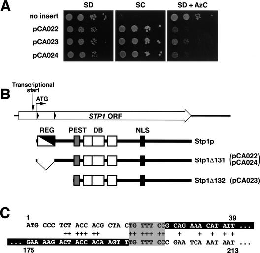
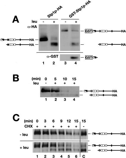
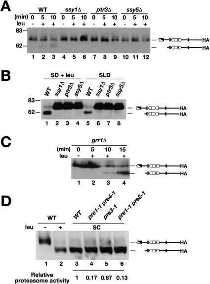
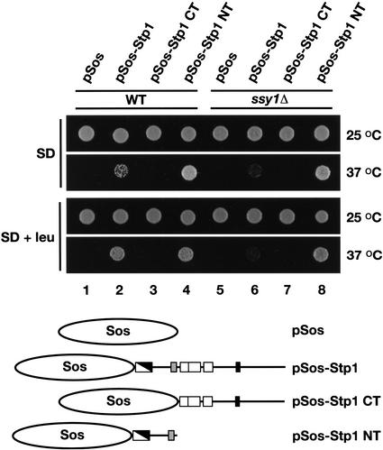
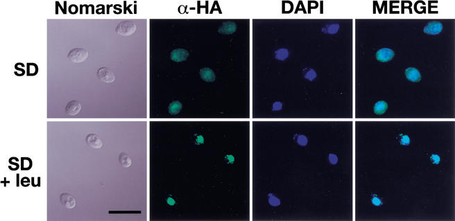
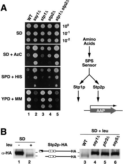
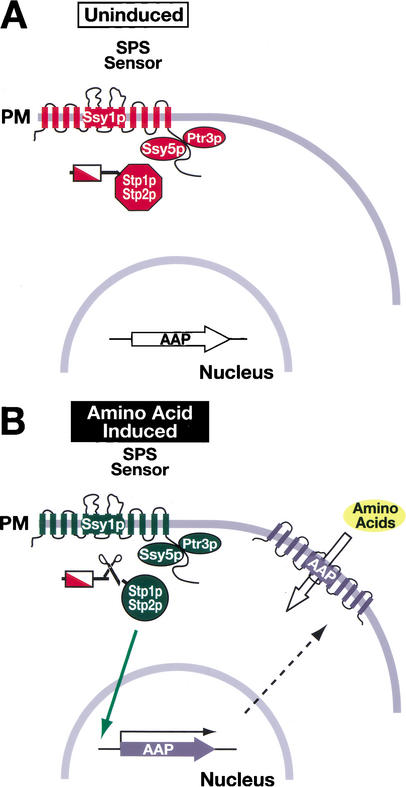
Yeast possess a plasma membrane sensor of external amino acids that functions as a ligand-activated receptor. This multimeric sensor, dubbed the SPS sensor, initiates signals that regulate the expression of genes required for proper amino acid uptake. Stp1p and Stp2p are transcription factors that bind to specific sequences within the promoters of SPS-sensor-regulated genes. These factors exhibit redundant and overlapping abilities to activate transcription. We have found that Stp1p and Stp2p are synthesized as latent cytoplasmic precursors. In response to extracellular amino acids, the SPS sensor induces the rapid endoproteolytic processing of Stp1p and Stp2p. The processing of Stp1p/Stp2p occurs independently of proteasome function and without the apparent involvement of additional components. The shorter forms of these transcription factors, lacking N-terminal inhibitory domains, are targeted to the nucleus, where they transactivate SPS-sensor target genes. These results define a completely unique and streamline metabolic control pathway that directly routes environmental signals initiated at the plasma membrane to transcriptional activation in the nucleus of yeast.


Figures

Figure 1
Figure 1
The dominant ASI13-1 (STP1Δ131) allele is an in-frame deletion that removes sequences encoding an inhibitory domain in the N terminus of Stp1p. (A) Phenotypic analysis of strain YMH119 transformed with plasmids pCA022 (STP1Δ131), pCA023 (STP1Δ132), and pCA024 (STP1Δ131). Transformants were spotted onto agar plates containing SD, SC, and SD containing AzC. Plates were incubated at 30°C for 3 d and photographed. (B) Schematic diagram of the STP1 gene. The start site of transcription is located within the ORF (Wang et al. 1992). The positions of the second ATG codon (translational start) and direct sequence repeats (black triangles) within STP1 are indicated. The proteins encoded by wild-type STP1 and the two dominant alleles STP1Δ131 and STP1Δ132 are depicted schematically. The regulatory domain, defined by the sequence between the repeats (REG, white/black diagonal box; amino acids 9–67), is absent in the constitutively active Stp1Δ131p. A putative nuclear localization signal (NLS, black box; amino acids 351–363), DNA-binding domain (DB, open boxes) with three putative zinc fingers (amino acids 160–187, 188–228, and 240–270), and a predicted PEST region (PEST, gray box; amino acids 129–147) are marked. (C) The sequences comprising the direct nucleotide repeats in STP1 are aligned (nucleotides 1–39 and 175–213, respectively). Matching base pairs are marked +, and the sequences removed in the STP1Δ131 deletion allele are indicated as white text within black boxes.
Figure 2
Figure 2
Characteristics of Stp1p processing. The inhibitory domain of Stp1p is removed in response to amino acids via an endoproteolytic processing event. (A) Immunoblotting of whole-cell extracts from strain CAY59 (stp1Δ) transformed with plasmid pCA047 (Stp1p–HA) or pCA073 (GST–Stp1p–HA) grown in SD or SD supplemented with leucine (SD + leu). Extracts were resolved on a 10% SDS-PAGE gel and serially immunoblotted with anti-HA (upper panel) and anti-GST (lower panel) antibodies. (B) Stp1p activation occurs rapidly. Immunoblotting of whole-cell extracts from strain CAY59 transformed with plasmid pCA047 (Stp1p–HA). Cells were pregrown in liquid SD, and at t = 0 the culture received an aliquot of leucine. At the times indicated, subsamples were removed and whole-cell extracts were prepared. (C) The proteolytic processing of Stp1p does not require de novo protein synthesis. Immunoblotting of whole-cell extracts from strain CAY59 transformed with plasmid pCA047 (Stp1p–HA). Cells were pregrown as in B; at t = 0 the culture was split into four equal volumes. These subcultures received an aliquot of cycloheximide (CHX, final concentration = 100 μg/mL) and leucine as indicated. The immunoreactive forms of Stp1p are schematically represented at their corresponding positions of migration.
Figure 3
Figure 3
The amino-acid-induced cleavage of Stp1p requires an operational SPS sensor and occurs independently of Grr1p and proteasome function. (A) Immunoblotting of whole-cell extracts from strains wild-type (WT, PLY126), ssy1Δ (HKY20), ptr3Δ (HKY31), and ssy5Δ (HKY77) cotransformed with plasmids pRS317 (LYS2) and pCA047 (Stp1p–HA). Cells were grown and induced with leucine as in Figure 2B. (B) Immunoblotting of whole-cell extracts from the strains grown in SD supplemented with leucine (SD + leu), or in SLD with leucine as the sole nitrogen source. After the preparation of extracts, proteins were resolved on 10% SDS-PAGE gels and immunoblotted with anti-HA antibody. (C) Immunoblotting of whole-cell extracts of CAY86 (grr1Δ) transformed with pCA047. Cells were grown and induced with leucine as in Figure 2B. (D) The migration of Stp1p–HA was analyzed in wild-type (WT, strain WCG4a) transformed with pCA047 and grown in SD before (lane 1) and after (lane 2) induction with leucine. Whole-cell extracts from WT (WCG4a, lane 3) and isogenic strains carrying mutations in the indicated proteasome components (pre1-1 pre4-1, pre3-1, and pre1-1 pre2-1, lanes 46, respectively) grown in SC (−ura) were prepared, and the migration of Stp1p–HA (pCA047) was examined by immunoblotting. The relative residual proteasome activity within these strains is indicated; proteasome activity was assessed by comparing the stability of model N-end rule substrates (Bachmair et al. 1986).
Figure 4
Figure 4
Stp1p associates with the plasma membrane. Prototrophic wild-type (WT, CAY187) and ssy1Δ (CAY189) strains carrying a temperature-sensitive cdc25 allele were transformed with plasmids pSos (pCA094), pSos-Stp1 (pCA093), pSos-Stp1 CT (pCA099), and pSos-Stp1 NT (pCA104). The proteins expressed from these plasmids are schematically presented. Transformants were selected and grown on solid SD incubated at 25°C. Cell suspensions were prepared in water (OD600 of 1), and equal aliquots of each suspension were spotted onto SD or SD containing leucine (SD + leu). Culture plates were incubated at 25°C or 37°C as indicated, and after 4 d the plates were photographed.
Figure 5
Figure 5
Proteolytically processed Stp1p is targeted to the nucleus. Indirect immunolocalization of full-length (upper panels) and proteolytically processed (lower panels) Stp1p was performed with anti-HA monoclonal antibodies. Strain CAY59 was transformed with pCA078 (pHXT7-STP1-HA) and grown in SD to an OD600 of 0.7. The culture was split into two equal aliquots, and leucine was added to one of the cultures. Both cultures were incubated shaking at 30°C for an additional 30 min, and the cells were fixed. (Panels, left to right) Cells viewed by Nomarski optics; α-HA monoclonal antibody-dependent Alexa Fluor 488 fluorescence; DAPI staining; and the MERGE of α-HA-dependent and DAPI fluorescence. (Lower left panel) Bar, 10 μm.
Figure 6
Figure 6
Stp1p and Stp2p exhibit overlapping and redundant function. (A) Wild-type (WT, CAY29), ssy1Δ (CAY91), stp1Δ (CAY59), stp2Δ (CAY119), and stp1Δ stp2Δ (CAY123) strains were grown on SD plates. Cells were resuspended in water, 10-fold serial dilutions were prepared, and aliquots of each dilution were applied to SD containing leucine (SD), SD containing leucine and AzC (SD + AzC), SPD containing toxic levels of histidine (SPD + HIS), and YPD containing MM (YPD + MM). In all instances synthetic media were supplemented with uracil. Plates were incubated at 30°C, and after 4 d plates were photographed. (B) SPS-sensor-initiated signals promote the proteolytic processing of Stp2p. Immunoblotting of whole-cell extracts from strain CAY119 transformed with pCA111 (Stp2p–HA) grown in SD or SD supplemented with leucine (SD + leu; left panel). Immunoblotting of whole-cell extracts from strains wild-type (WT, PLY126), ssy1Δ (HKY20), ptr3Δ (HKY31), and ssy5Δ (HKY77) cotransformed with plasmids pRS317 (LYS2) and pCA111 (Stp2p–HA) grown in SD supplemented with leucine (SD + leu; right panel). The immunoreactive forms of Stp2p are schematically represented at their corresponding positions of migration.
Figure 7
Figure 7
Model of the SPS-sensor-dependent activation of Stp1p and Stp2p. (A) In cells grown in the absence of inducing amino acids, the SPS sensor of extracellular amino acids is present in the plasma membrane (PM) in its preactivation conformation (Forsberg and Ljungdahl 2001a). The transcription factors Stp1p and Stp2p are synthesized as inactive precursors that localize to the cytosol. The transcription of SPS-sensor-regulated genes, for example, AAPs, occurs at basal levels. Consequently, there are correspondingly low levels of AAPs in the PM, and cells exhibit low rates of amino acid uptake. (B) In the presence of inducing amino acids, the SPS sensor is activated, leading to the endoproteolytic processing of Stp1p and Stp2p. The shorter, activated forms of Stp1p and Stp2p, lacking the inhibitory domains located within their N termini, are targeted to the nucleus (solid arrow), where they function to induce the transcription of SPS-sensor-regulated genes. The increased transcription of AAP genes results in a comcomitant increase in AAPs in the PM (translation and movement to the PM are represented by the dashed arrow), and cells exhibit induced rates of amino uptake.

References

    1. André B. An overview of membrane transport proteins in Saccharomyces cerevisiae. Yeast. 1995;11:1575–1611. - PubMed
    1. Aronheim A, Zandi E, Hennemann H, Elledge SJ, Karin M. Isolation of an AP-1 repressor by a novel method for detecting protein–protein interactions. Mol Cell Biol. 1997;17:3094–3102. - PMC - PubMed
    1. Bachmair A, Finley D, Varshavsky A. In vivo half-life of a protein is a function of its amino-terminal residue. Science. 1986;234:179–186. - PubMed
    1. Barnes D, Lai W, Breslav M, Naider F, Becker JM. PTR3, a novel gene mediating amino acid-inducible regulation of peptide transport in Saccharomyces cerevisiae. Mol Microbiol. 1998;29:297–310. - PubMed
    1. Bernard F, André B. Genetic analysis of the signalling pathway activated by external amino acids in Saccharomyces cerevisiae. Mol Microbiol. 2001a;41:489–502. - PubMed

Publication types

MeSH terms