. 2002 Dec 15;16(24):3236-52.
doi: 10.1101/gad.239802.

ORC and the intra-S-phase checkpoint: a threshold regulates Rad53p activation in S phase

Affiliations

ORC and the intra-S-phase checkpoint: a threshold regulates Rad53p activation in S phase

Kenji Shimada et al. Genes Dev. .

Abstract

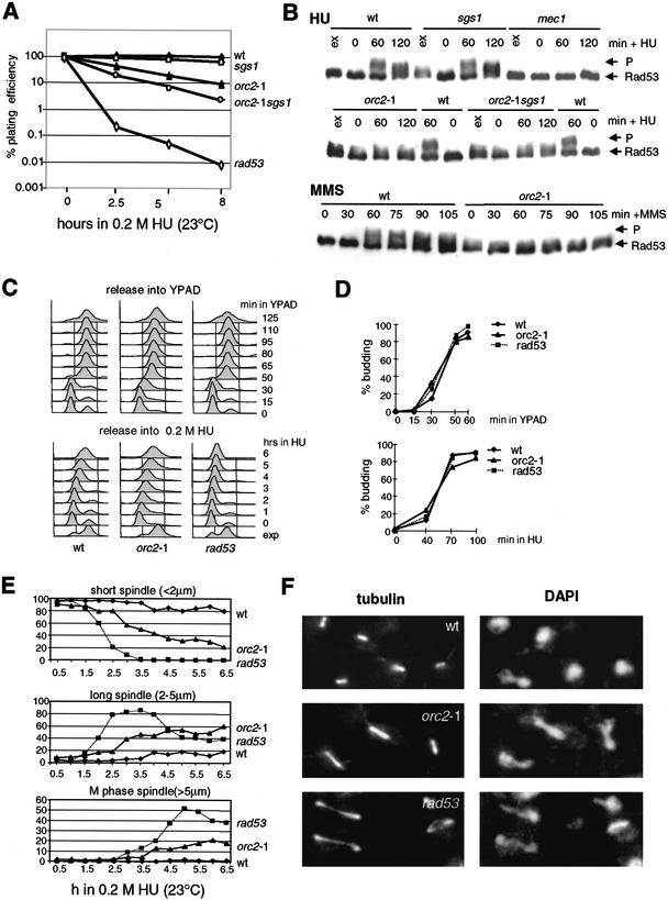
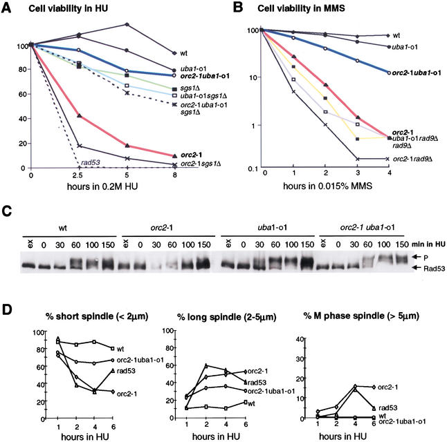
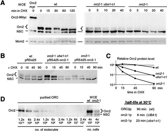
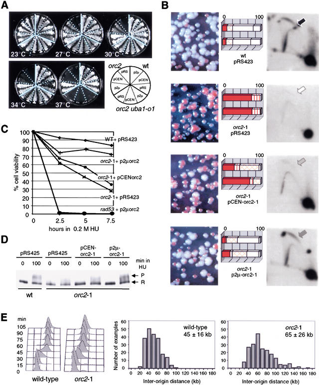
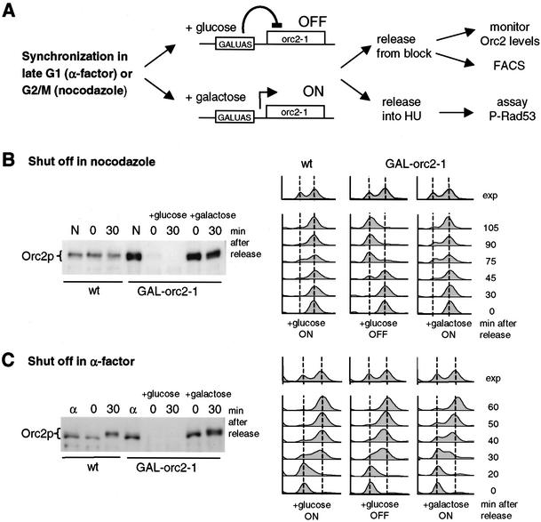
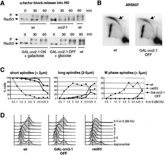
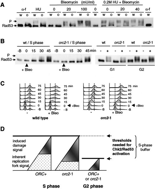
The intra-S-phase checkpoint in yeast responds to stalled replication forks by activating the ATM-like kinase Mec1 and the CHK2-related kinase Rad53, which in turn inhibit spindle elongation and late origin firing and lead to a stabilization of DNA polymerases at arrested forks. A mutation that destabilizes the second subunit of the Origin Recognition Complex, orc2-1, reduces the number of functional replication forks by 30% and severely compromises the activation of Rad53 by replication stress or DNA damage in S phase. We show that the restoration of the checkpoint response correlates in a dose-dependent manner with the restoration of pre-replication complex formation in G1. Other forms of DNA damage can compensate for the reduced level of fork-dependent signal in the orc2-1 mutant, yet even in wild-type cells, the amount of damage required for Rad53 activation is higher in S phase than in G2. Our data suggest the existence of an S-phase-specific threshold that may be necessary to allow cells to tolerate damage-like DNA structures present at normal replication forks.


Figures

Figure 1
Figure 1
orc2-1 cells are defective in S-phase checkpoint. (A) orc2-1 cells are hypersensitive to HU. Isogenic strains carrying the orc2-1 mutation, a deletion of sgs1, both mutations, or rad53-11 (mec2-1) were synchronized by α-factor, then released into 0.2 M HU for the indicated times at 23°C. Washed cells were plated in triplicate on YPAD, and plating efficiency was determined after 3 d. Values for each strain are normalized to survival at time 0. ♦, wild-type (GA-180); ▴, orc2-1 (GA-1254); □, sgs1∷TRP1 (GA-734); ○, orc2-1 sgs1∷TRP1 (GA-1387); ⋄, rad53-11 (mec2-1; GA-1230). (B) Exponentially growing cultures (ex) were blocked in G1 with α-factor and released in 0.2 M HU (+ HU) or 0.015% MMS (+ MMS) for the indicated times at 23°C. Western blots of whole-cell extracts monitor phosphorylation of Rad53-Myc with the 9E10 Mab. P indicates the fully phosphorylated form of Rad53p. Isogenic strains were as follows: wild-type (wt, GA-1829, A364a background); sgs1∷LEU2 (sgs1, GA-1830); mec1-1 sml1 (mec1, GA-1048); orc2-1 (GA-1831); orc2-1 sgs1∷LEU2 (orc2-1 sgs1, GA-1832). (C) Wild-type, orc2-1 and rad53-11 strains as in A were cultured and blocked with α-factor at 23°C. Cells were released in YPAD with or without 0.2 M HU at 23°C, and samples analyzed by FACS at the indicated times. (D) Bud emergence was scored microscopically on 100 cells of cultures examined in C. (E) Isogenic strains carrying an integrated GFP–TUB1 fusion (tubulin) were blocked with α-factor and released into 0.2 M HU. At the indicated times, spindle length was monitored by live fluorescence microscopy. For each genotype and each time point, 100 cells were measured. Strains used are as follows: wild-type (♦, GA-1535), orc2-1 (▴, GA-1533), and rad53 (mec2-1; ■, GA-1499). (F) The strains used in A were synchronized by α-factor, then released into 0.2 M HU for 5 h at 23°C. Tubulin is visualized with anti-TAT1 antibody and DNA with DAPI.
Figure 2
Figure 2
The S-phase checkpoint response is restored in an orc2-1 uba1-o1 double mutant. (A,B) The uba1-o1 mutation suppresses the hypersensitivity of orc2-1 cells to HU (A) and MMS (B). Viability assays were performed as in Figure 1A after indicated times in 0.2 M HU or 0.015% MMS. Isogenic strains used in A and B were as follows: wild-type (GA-180); sgs1∷TRP1 (GA-734); orc2-1 (GA-1254); uba1-o1 (GA-1256); orc2-1 uba1-o1 (GA-463); orc2-1 sgs1∷TRP1 (GA-1387); uba1-o1 sgs1∷TRP1 (GA-1388); orc2-1 uba1-o1 sgs1∷TRP1 (GA-1389); rad53 (mec2-1; GA-1230); rad97∷LEU2 (GA-1148), orc2-1 rad9∷LEU2 (GA-1860); uba1-o1 rad9∷LEU2 (GA-1861). (C) Rad53p phosphorylation in response to HU is restored in an orc2-1 uba1-o1 double mutant (see Fig. 1B). Strains were as follows: wild-type (GA-1835); orc2-1 (GA-1836); uba1-o1 (GA-1837); orc2-1 uba1-o1 (GA-1838). (D) The uba1-o1 mutation restores the cell cycle arrest response to HU in orc2-1 cells. Cells synchronized in G1 as in A were released into YPAD + 0.2 M HU. Spindle elongation was scored as in Figure 1F. The drop in long spindles in the rad53 strain at 6 h reflects entry into the next cell cycle.
Figure 3
Figure 3
The highly labile protein orc2-1p is partially stabilized by the uba1-o1 mutation. (A) Cycloheximide was added to exponentially growing cells and total protein extracts were prepared at indicated times. Western blots were sequentially probed with goat anti-MCM2 (Santa Cruz Antibodies) or affinity-purified anti-Orc2p antibody. The brace indicates to a doublet of Orc2p-specific bands (the upper band being phosphorylated); NSC indicates a nonspecific cross-reaction with a cytoplasmic protein that serves as an internal control for loading. Strains used were orc27∷ORC2-9Myc (GA-893); wild-type (GA-180); orc2-1 uba1-o1 (GA-463); orc2-1 (GA-1254). (B) Isogenic orc2-1 and orc2-1 uba1-o1 cells transformed with a multicopy plasmid expressing mutant orc2-1p (pRS425-orc2-1) and wild-type cells with the empty vector, were treated with cycloheximide (CHX) at 30°C, and whole-cell extracts were prepared at the indicated times. Orc2p protein was revealed as in A. (C) Orc2p protein signals in B were quantified by the AIDA program (Fuji PhosPhorimager) and normalized to the lower nonspecific band (NSC). Relative intensity is plotted as a function of time after CHX addition and the half-life of Orc2p (t1/2) was calculated. (D) Decreasing amounts of purified recombinant Baculovirus-expressed yeast ORC complex (gift of Dr. S. Bell), and total protein extracts equivalent to 4 × 105 cells from isogenic wild-type and orc2-1 cells grown at 23°C, were analyzed by Western blot using affinity-purified anti-Orc2 antibodies.
Figure 4
Figure 4
Ectopic expression of the orc2-1 mutant protein suppresses orc2 defects in a dosage-dependent manner. (A) Two independent transformants of orc2-1, orc2-1 uba1-o1, and wild-type cells carrying with p2μ (pRS425-orc2-1), pCEN (pRS415-orc2-1) or pRS425 (labeled pRS) were streaked on SD-leu and cultured at the indicated temperature (see diagram). (B) The orc2-1 strain was cotransformed with pRS415-ADE2 (carrying ADE2 and CEN6/H4ARS) and pRS423-orc2-1 (p2μ-orc2-1), or pRS413-orc2-1 (pCEN-orc2-1), and both wild-type and orc2-1 cells were cotransformed with pRS415-ADE2 and pRS423. Transformants were first selected on SD-leu-his, then plated on SD-his containing limiting amounts of adenine for 3–4 d at 23°C. The rate of pRS415-ADE2 loss scored by the level of red pigment reflects the initiation efficiency and was scored for 100–200 colonies from two independent transformants. Values are presented as percentage of total colony number. Solid box, all red colony; vertical-lined box, >80% red; cross-hatched box, 20%–80% red; diagonal-lined box, <20% red; open box, white. NcoI-digested genomic DNA from each transformant was probed for the genomic ARS1 after two-dimensional-gel electrophoresis (see Materials and Methods). Arrows identify bubble arcs (i.e., initiation events). (C) Isogenic wild-type, orc2-1 and rad53 strains were transformed with pRS423, whereas rad53 and orc2-1 strains were transformed with p2μ-orc2-1 (pRS423-orc2-1) or pCEN-orc2-1 (pRS413-orc2-1). Transformants cultured in SD-his at 23°C were scored for cell viability as in Figure 1A. (D) Isogenic wild-type and orc2-1 strains transformed with vector or orc2-1 expressing plasmids (A–C) were cultured in SD-leu at 23°C. Western analysis of Rad53-myc phosphorylation was performed (see Fig. 1) and Rad53p phosphorylation levels in orc2-1 cells after 100 min in HU were 49% (pRS425), 63% (pCEN-orc2-1), and 97% (p2μ-orc2-1) of the wild-type levels. (E) DNA combing and quantitation of the distances between stretches of BrdU incorporation were performed as described in Materials and Methods by use of wild-type (E-1000) and orc2-1 cells (E-1313) cultured at 24°C. The inter-origin distances (kilobase) for 208 origins are plotted, and the mean and S.D. indicated. Cell cycle progression through S phase was monitored by FACS analysis at 24°C.
Figure 5
Figure 5
The integrated GAL1UASorc2-1 construct allows rapid depletion of orc2-1p in synchronized cells. (A) After integration of Gal1UAS upstream of the mutant orc2-1 gene (GAL1UAS∷orc2-1), orc2-1p levels are slightly elevated on galactose and the cells exhibit wild-type growth rates (B,C). Replacing galactose with glucose suppresses expression and all detectable orc2-1p disappears within 60 min. (B,C) Wild-type (GA-180) and GAL1UASorc2-1 cells (GA-1680) were cultured in YPA-2% galactose at 30°C, and cells were arrested at G2/M with nocodazole (7.5 μg/mL, B) for 70 min or at G1 with α-factor for 60 min (C). Thereafter, the wild-type and half of the GAL1UASorc2-1 cells were shifted for 1 h to 2% glucose medium with nocodazole or pheromone to allow depletion of orc2-1p. Cells were then released from the nocodazole or α-factor block into either glucose or galactose, as indicated. Samples were taken for Western blots with anti-Orc2p at 0 or 30 min (minutes after release), or for FACS analysis at 30–105 min (B) and 20–60 min (C).
Figure 6
Figure 6
The S-phase checkpoint requires ORC2 function in G1, but not in S phase. (A) Congenic wild-type (GA-1682), GAL1UASorc2-1 (GA-1681), and orc2-1 (GA-1836) strains were cultured in YPA-2% galactose (YPAG) at 23°C. After synchronization in α-factor, wild-type, orc2-1, and half of the GAL1UASorc2-1 (GAL∷orc2-1 OFF) cells were switched to YPAD + α-factor, whereas the other half of the GAL1UASorc2-1 culture was kept on YPAG + α-factor (GAL∷orc2-1 ON). After 60 min at 30°C, cultures were washed and released from pheromone into 0.2 M HU in YPAD or YPAG at 30°C. Rad53p phosphorylation was monitored at 0, 30, and 60 min after release. (B) Two-dimensionl gel analysis of replication intermediates at the genomic origin ARS607, based on genomic DNA prepared from wild-type and GAL∷orc2-1 OFF cells released from α-factor into HU for 60 min. PstI–ClaI-digested genomic DNA was probed as described in Materials and Methods. (C,D) Wild-type (GA-1535), rad53 (GA-1499), and GAL1UASorc2-1 (GA-1780) cells were synchronized by α-factor in YPAG at 30°C. After depletion of orc2-1p during α-factor block (A), cells were released into YPAD + 0.2 M HU, and aliquots were taken for spindle elongation analysis and FACS (D) as in Figure 1C–E. At 4–5 h, rad53 cells enter the next cell cycle.
Figure 7
Figure 7
Combined damage reaches the threshold for intra-S-phase checkpoint activation. (A) Wild-type (w, GA-1829) and orc2-1 (o, GA-1831) strains were cultured at 23°C and synchronized in G1 with α-factor (α-f). After release into YPAD without HU, early S-phase cells were exposed to 0, 20, or 100 mU/mL bleomycin for 30 min, and then analyzed for Rad53p phosphorylation. Samples to the right were identical, except that cells were first released into YPAD + 0.2 M HU for 50 min (HU), prior to bleomycin addition (0, 20, or 40 mU/ml for 30 min). (B) For the analysis of Rad53p activation in S-phase, wild-type (wt, GA-1835) and orc2-1 (GA-1836) cells were blocked with α-factor at 23°C and released into YPAD. The functional equivalent of 20 mU/mL bleomycin was added after 20 min, and Rad53-myc phosphorylation (P) was monitored at the indicated times. For G1- or G2-phase cells, Rad53 activation was analyzed in α-factor-arrested or nocodazole-arrested cells, either with (+) or without (−) exposure to the same level of bleomycin for 30 min (right). Batch-specific variations in milliunits of bleomycin are normalized by functional assays. (C) S-phase progression with (+Bleo) or without (−) 20 mU/mL bleomycin was determined by FACS on samples from B, left. (D) We propose that the Chk2 (Rad53p)-dependent checkpoint in S phase, unlike that in G2 phase, has a buffer zone that tolerates a certain level of activating signal to allow normal fork progression. The signal arising from natural replication fork pausing is a likely source of background signal (hatched region) that can be complemented by signals arising from fork arrest (DNA pol ɛ and/or Sgs1-dependent signals) or DNA damage (Rad9- or Rad24-dependent signals), to reach an activation threshold. In orc2-1 cells, replication fork number is lower than in wild-type cells, more damage-induced signal is necessary to achieve Rad53p activation.

References

    1. Alcasabas AA, Osborn AJ, Bachant J, Hu F, Werler PJ, Bousset K, Furuya K, Diffley JF, Carr AM, Elledge SJ. Mrc1 transduces signals of DNA replication stress to activate Rad53. Nat Cell Biol. 2001;3:958–965. - PubMed
    1. Bell SP. The origin recognition complex: From simple origins to complex functions. Genes & Dev. 2002;16:659–672. - PubMed
    1. Blow JJ, Gillespie PJ, Francis D, Jackson DA. Replication origins in Xenopus egg extract are 5–15 kilobases apart and are activated in clusters that fire at different times. J Cell Biol. 2001;152:15–25. - PMC - PubMed
    1. Deshpande AM, Newlon CS. DNA replication fork pause sites dependent on transcription. Science. 1996;272:1030–1033. - PubMed
    1. Diffley JF. DNA replication: building the perfect switch. Curr Biol. 2001;11:R367–R370. - PubMed

Publication types

MeSH terms