. 2003 Jan 7;100(1):342-6.
doi: 10.1073/pnas.0135434100. Epub 2002 Dec 26.

A macromolecular complex of beta 2 adrenergic receptor, CFTR, and ezrin/radixin/moesin-binding phosphoprotein 50 is regulated by PKA

Affiliations

A macromolecular complex of beta 2 adrenergic receptor, CFTR, and ezrin/radixin/moesin-binding phosphoprotein 50 is regulated by PKA

Anjaparavanda P Naren et al. Proc Natl Acad Sci U S A. .

Abstract

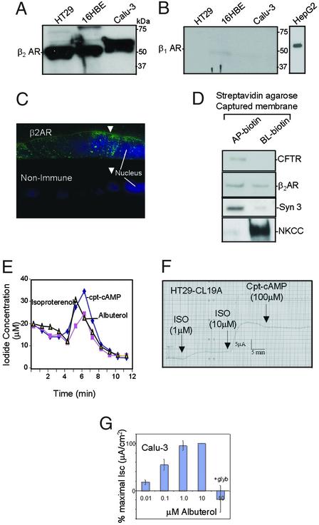
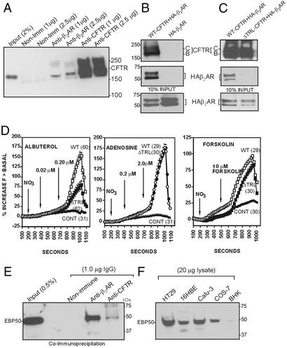
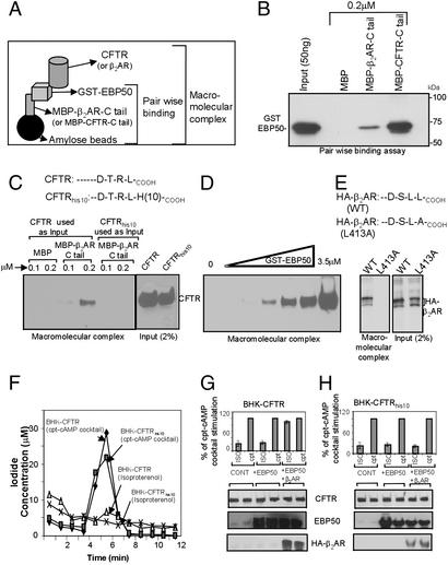
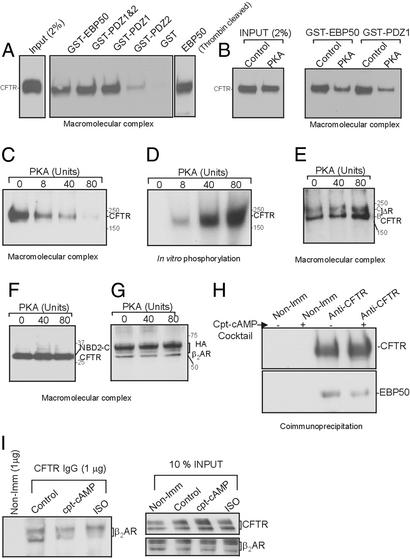
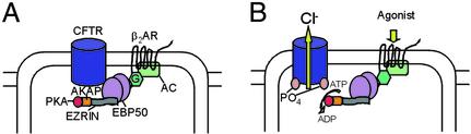
It has been demonstrated previously that both the cystic fibrosis transmembrane conductance regulator (CFTR) and beta(2) adrenergic receptor (beta(2)AR) can bind ezrinradixinmoesin-binding phosphoprotein 50 (EBP50, also referred to as NHERF) through their PDZ motifs. Here, we show that beta(2) is the major adrenergic receptor isoform expressed in airway epithelia and that it colocalizes with CFTR at the apical membrane. beta(2)AR stimulation increases CFTR activity, in airway epithelial cells, that is glybenclamide sensitive. Deletion of the PDZ motif from CFTR uncouples the channel from the receptor both physically and functionally. This uncoupling is specific to the beta(2)AR receptor and does not affect CFTR coupling to other receptors (e.g., adenosine receptor pathway). Biochemical studies demonstrate the existence of a macromolecular complex involving CFTR-EBP50-beta(2)AR through PDZ-based interactions. Assembly of the complex is regulated by PKA-dependent phosphorylation. Deleting the regulatory domain of CFTR abolishes PKA regulation of complex assembly. This report summarizes a macromolecular signaling complex involving CFTR, the implications of which may be relevant to CFTR-dysfunction diseases.


Figures

Figure 1
Figure 1
β2AR is expressed at the apical surface of airway epithelial cells and can physically and functionally interact with CFTR. (A) Western blot analysis with affinity-purified β2AR antibodies (Santa Cruz Biotechnology; 1 μg/ml) shows that β2AR is expressed in HT29-CL19A (colonic epithelial), 16HBE14o− (human bronchial epithelial), and calu-3 (serous gland epithelial) cell lysates (100 μg per lane). (B) β1AR is not detected in these cells using affinity-purified β1AR antibody (Santa Cruz Biotechnology; 1 μg/ml; HepG2 cell lysate was used as positive control). (C) Immunofluorescence localization of β2AR at the apical membrane in calu-3 cells grown on a permeable supports. Polarized monolayers were fixed and stained after 8 days in culture as described (20). AP, apical; BL, basolateral. Arrows indicate the apical membrane. The nucleus was stained with Hoechst reagent (20). (D) Colocalization of CFTR and β2AR in apical membranes of polarized calu-3 cells (see Materials and Methods). (E) Iodide effluxes were measured by using calu-3 monolayers (n = 2) according to described methods (21). Cells were activated after 4 min with cpt-cAMP mixture [200 μM cpt-cAMP/10 μM forskolin/1 mM 3-isobutyl-1-methylxanthine (IBMX)], isoproterenol (50 μM), or albuterol (100 μM). (F) Activation of dose-dependent anion secretion by adding isoproterenol to the apical surfaces of polarized HT29-CL19A cells mounted in an Ussing chamber (23). The currents could not be potentiated further by cpt-cAMP. (G) Activation of glibenclamide-sensitive anion secretion by β2AR agonist added to the apical side of calu-3 monolayers mounted in Ussing chambers (area = 0.33 cm2; n = 4; ref. 23). Cells were treated with apical albuterol (0–10 μM), followed by apical glybenclamide (200 μM).
Figure 2
Figure 2
Physical and functional interaction between CFTR and β2AR is mediated by the PDZ motif of CFTR. (A) CFTR coimmunoprecipitates with β2AR from calu-3 cells. Ten confluent 100-mm dishes were lysed in PBS–0.2% Triton X-100, and CFTR was immunoprecipitated by using a polyclonal CFTR antibody (raised against amino acid residues 521–828) cross-linked to protein A/G beads (11). (B) Coimmunoprecipitation of CFTR and β2AR in Cos-7 cells expressing recombinant CFTR and β2AR (vaccinia expression system; ref. 35). The blot was cut at an appropriate position and probed by using the monoclonal antibodies GA-1 (recognizes amino acids 1430–1460 of CFTR; Top) and anti-HA tag mAb (Sigma; Middle). (C) Deletion of the PDZ motif (ΔTRL-CFTR) abolished the interaction of CFTR with HA-β2AR. The vaccinia expression system generated approximately equal steady-state levels of mature (band C) and immature (band B) CFTR. (D) Halide transport by ΔTRL-CFTR expressed in Cos-7 cells is reduced on stimulation by β2AR agonist (albuterol, Left), compared with WT-CFTR (Left). In contrast, deletion of the TRL motif of CFTR had no effect on halide transport stimulated by adenosine receptor agonist, shown in Center, and forskolin, shown at Right, compared with WT-CFTR. There was no significant difference in the average basal level of halide transport in the absence of stimulation of WT-CFTR vs. ΔTRL-CFTR. The total protein levels were not altered (data not shown). Fluorescence of the halide-sensitive indicator dye (SPQ) was assayed as described (22). (E) Cross-linked β2AR IgG (1 μg), polyclonal CFTR IgG (1 μg), or nonimmune IgG (1 μg, Santa Cruz Biotechnology) was used to immunoprecipitate 16HBE14o− cell lysate (PBS–0.2% Triton X-100). The blots were probed by using monoclonal EBP50 antibody (Transduction Laboratories; 2 μg/ml). (F) Western blot analysis of lysates of various cell lines using affinity-purified EBP50 polyclonal antibody (see Materials and Methods). The protein was detected (EBP50) in all cell lines tested.
Figure 3
Figure 3
Macromolecular complex of CFTR and β2AR is mediated by EBP50. (A) Pictorial representation of the in vitro macromolecular assembly (see Materials and Methods). (B) C-terminal tail of CFTR and β2AR interacted directly with EBP50 as determined by pair-wise binding assay. MBP-fusion proteins (0.02 μM) were used to bind 0.001 μM GST-EBP50 for 60 min at 22°C in lysis buffer. The complex was pulled down by amylose beads (20 μl) and washed twice with the same buffer. The complex was blotted and probed by using anti-EBP50 monoclonal antibody. (C) Macromolecular complex of β2AR, EBP50, and CFTR. A complex between MBP-β2AR-C tail (0.5 μM) and GST-EBP50 (0.5 and 1.0 μM) was formed in lysis buffer (200 μl final volume) as described above, washed with 1 ml of the same buffer, and mixed with 1 ml of BHK cell lysate (PBS–0.2% Triton X-100) containing CFTR or CFTRhis10. The proteins were bound for 3 h at 4°C, washed twice with the same buffer, and subjected to blotting by using GA1 mAb (11). BHK cells stably expressing CFTR contained mainly the complex glycosylated mature “band C” form. The inputs are shown at Right. (D) Dose-dependent complex formation with increasing amounts of EBP50. Saturation was not observed up to 3.5 μM. (E) Formation of a macromolecular complex MBP-CFTR-C tail (0.5 μM), GST-EBP50 (1.0 μM), and HA-β2AR (WT and mutant; L413A). (F) BHK cells expressing CFTR or CFTRhis10 did not respond to isoproterenol (100 μM) or albuterol (data not shown). These cells responded to cpt-cAMP mixture. (G) BHK-CFTR cells were transiently transfected with 1 μg of EBP50 with or without HA-β2AR cDNA (Lipofectamine 2000; see Materials and Methods) and subjected to iodide efflux by using isoproterenol (ISO) or the cpt-cAMP (cpt) mixture. The data are presented as the percentage of cpt-cAMP mixture stimulation (at 5-min time points). (H) BHK-CFTRhis10 cells were transiently transfected with 1 μg of EBP50 with or without HA-β2AR cDNA and subjected to iodide efflux as described above.
Figure 4
Figure 4
Association of CFTR with β2AR and EBP50 complex is regulated by PKA. (A) Macromolecular complex formation between MBP-β2AR-C tail (1 μM) and CFTR by using various EBP50 constructs (1 μM). The assay is described in Materials and Methods. (B) Effect of PKA on macromolecular complex formation. ATP (2 mM), Mg (2 mM), and PKA (80 units) were added to the cell lysate, and the complex was formed by stepwise assembly as described (see Materials and Methods). (C) A dose-dependent inhibitory effect of PKA (0–80 units) on macromolecular complex formation is shown. (D) In vitro phosphorylation of CFTR was performed in parallel, verifying that the protein was being appropriately phosphorylated. (E) Deleting most of the R domain (ΔR-CFTR; ref. 32) eliminated the inhibitory effects of PKA on the complex formation. The vaccinia expression system was used to express ΔR-CFTR, which yielded equal amounts of immature and mature protein (bands B and C). (F) NBD-2-C tail of CFTR (amino acids 1210–1480) also eliminated the PKA inhibitory effects on the macromolecular complex formation (vaccinia expression system was used to generate the protein). (G) PKA phosphorylation did not affect β2AR in the complex of MBP-CFTR-C tail (1 μM) and GST-EBP50 (1 μM). In vitro assembly of the complex is described (see Materials and Methods). Vaccinia expression system was used to generate HA-β2AR. (H) Coimmunoprecipitation of CFTR and EBP50 from calu-3 cells treated with or without cpt-cAMP mixture for 10 min at 37°C. cpt-cAMP treatment diminishes EBP50 binding during coimmunoprecipitation. (I) Coimmunoprecipitation of CFTR and β2AR from COS-7 cells (expressing recombinant CFTR and HA-β2AR) treated with agonists for 10 min at 37°C.
Figure 5
Figure 5
A model of CFTR-EBP50-β2AR signaling. CFTR, β2AR, and EBP50 can exist as a complex at the apical surface of epithelial cells. G protein (G) can be associated with β2AR (data not shown; ref. 33) and protein kinase A (PKA) anchored to AKAP via ezrin (10) and is likely to be in the complex. On agonist activation of the receptor, adenylate cyclase is stimulated through the Gs pathway (33), leading to an increase in highly compartmentalized cAMP. This increased local concentration of cAMP leads to the activation of PKA, which is in close proximity to CFTR (36), leading to a compartmentalized and specific signaling of the channel. Phosphorylation disrupts the complex, leading to the receptor-based activation of CFTR.

Similar articles

Cited by

References

    1. Welsh M J, Tsui L-C, Boat T F, Beaudet A L. In: The Metabolic and Molecular Basis of Inherited Diseases: Membrane Transport Systems. Scriver C, Beaudet A L, Sly W S, Valle D, editors. Vol. 3. New York: McGraw–Hill; 1995. pp. 3799–3876.
    1. Gadsby D C, Nairn A C. Adv Second Messenger Phosphoprotein Res. 1999;33:79–106. - PubMed
    1. Kunzelmann K. News Physiol Sci. 2001;16:167–170. - PubMed
    1. Matsui H, Davis C W, Tarran R, Boucher R C. J Clin Invest. 2000;105:1419–1427. - PMC - PubMed
    1. Chan H C, Fong S K, So S C, Chung Y W, Wong P Y. J Physiol. 1997;501:517–525. - PMC - PubMed

Publication types

MeSH terms