. 2003 Jan 7;100(1):131-6.
doi: 10.1073/pnas.0135855100. Epub 2002 Dec 26.

Identification of an intracellular receptor for lysophosphatidic acid (LPA): LPA is a transcellular PPARgamma agonist

Affiliations

Identification of an intracellular receptor for lysophosphatidic acid (LPA): LPA is a transcellular PPARgamma agonist

Thomas M McIntyre et al. Proc Natl Acad Sci U S A. .

Erratum in

  • Proc Natl Acad Sci U S A. 2003 Feb 18;100(4):2163

Abstract

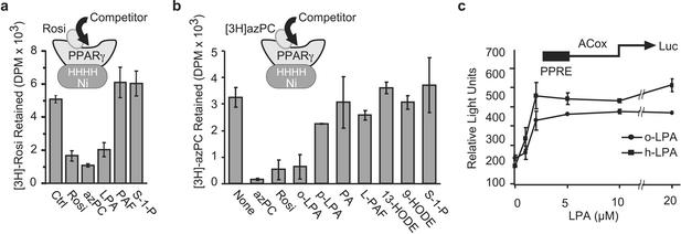
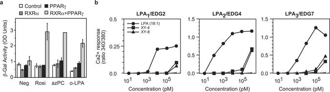
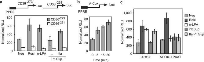
Lysophosphatidic acid (LPA) is a pluripotent lipid mediator acting through plasma membrane-associated LPA(x) receptors that transduce many, but not all, of its effects. We identify peroxisome proliferator-activated receptor gamma (PPARgamma) as an intracellular receptor for LPA. The transcription factor PPARgamma is activated by several lipid ligands, but agonists derived from physiologic signaling pathways are unknown. We show that LPA, but not its precursor phosphatidic acid, displaces the drug rosiglitazone from the ligand-binding pocket of PPARgamma. LPA and novel LPA analogs we made stimulated expression of a PPAR-responsive element reporter and the endogenous PPARgamma-controlled gene CD36, and induced monocyte lipid accumulation from oxidized low-density lipoprotein via the CD36 scavenger receptor. The synthetic LPA analogs were effective PPARgamma agonists, but were poor ones for LPA(1), LPA(2), or LPA(3) receptor transfected cells. Transfection studies in yeast, which lack nuclear hormone and LPA(x) receptors, show that LPA directly activates PPARgamma. A major growth factor of serum is LPA generated by thrombin-activated platelets, and media from activated platelets stimulated PPARgamma function in transfected RAW264.7 macrophages. This function was suppressed by ectopic LPA-acyltransferase expression. LPA is a physiologic PPARgamma ligand, placing PPARgamma in a signaling pathway, and PPARgamma is the first intracellular receptor identified for LPA. Moreover, LPA produced by stimulated plasma platelets activates PPARgamma in nucleated cells.


Figures

Figure 1
Figure 1
LPA is a PPARγ ligand and agonist. LPA competes for [3H]rosiglitazone (a) or [3H]azPC (b) binding to immobilized PPARγ. His6-PPARγ1 was immobilized on Ni+ beads and incubated with [3H]rosiglitazone or [3H]azPC in the presence or absence of a 20-fold molar excess of the stated lipids. Rosi, rosiglitazone; o-LPA, oleoyl LPA; p-LPA, palmitoyl LPA; PA, phosphatidic acid; PAF, 1-O-hexadecyl-2-acetyl-sn-glycero-3-phosphocholine; L-PAF, 1-O-hexadecyl-sn-glycero-3-phosphocholine; HODE, hydroxyoctadecadienoic acid; S-1-P, sphingosine 1-phosphate. (c) LPA induces a concentration-dependent increase in PPRE-luciferase reporter expression. RAW 264.7 monocytic cells were transfected with rat acyl-CoA oxidase PPRE-luciferase and SV40-β-galactosidase reporter plasmids. The transfected cells were treated with the stated amount of oleoyl LPA (o-LPA) or sn-1-hexadecyl LPA (h-LPA) for 16 h before the relative amounts of luciferase and β-galactosidase were determined. All luciferease data are reported as relative light units after normalization for transfection efficiency with β-galactosidase.
Figure 2
Figure 2
Intracellular LPA acyltransferase expression blocks LPA stimulation of a PPRE reporter, and novel nonacylatable LPA analogs are PPAR agonists. (a) Structure of: XY4, 1,1-difluorodeoxy-(2R)-oleoyl-sn-glycero-3-phosphate; XY8, 1-palmitoyl-(2R)-fluorodeoxy-sn-glycero-3-phosphate; (Left) translocase-3 Tris[2-(2-naphthalenesulfonamido)ethyl]amine interacting with oleoyl LPA. (b) A small organic cationic translocase enhances exogenous LPA stimulation of a PPRE reporter. RAW264.7 cells were transfected with an acyl-CoA oxidase-PPRE reporter and treated with 5 μM rosiglitazone, oleoyl LPA, or an sn-2 (XY4) or sn-1 (XY8) LPA analog. Some wells were additionally treated with 35 μM translocase-3. Normalized luciferase expression was determined as before. (c) LPA acyltransfersase suppresses LPA, but not nonacylatable LPA analog, stimulation of a PPRE reporter. RAW264.7 cells were transfected with acyl-CoA oxidase-PPRE and SV40-β-galactosidase reporters, and, for some cells, an LPA acyltransferase (LPAAT) pcDNAI expression construct before their response to the stated agonist was determined as above.
Figure 3
Figure 3
LPA is a direct PPARγ agonist. (a) LPA stimulates PPRE reporter transcription in S. cerevisiae after ectopic expression of PPARγ and RXR. S. cerevisiae was selected to stably express a PPRE-β-galactosidase reporter alone or the reporter and RXR, PPARγ, or both PPARγ and RXR. These cells were then incubated with the stated agonist for 16 h before the amount of β-galactosidase produced was quantified. (b) XY4 and XY8 are ineffective LPA1, LPA2, or LPA3 agonists. Sf9 cells were stably transfected with one of the three LPAx receptors, loaded with the Ca2+-sensitive dye fura-2, and then stimulated with the stated amount of LPA, XY4, or XY8 before changes in intracellular Ca2+ levels were determined from the ratio of fluorescence after excitation at two wavelengths.
Figure 4
Figure 4
LPA stimulates lipid accumulation, CD36 expression, and oxidized LDL uptake through a PPAR-responsive element. (a) LPA stimulates monocyte uptake of oxidized LDL. Freshly elutriated human monocytes were allowed to interact with an anti-ICAM3-coated well, which leads to rapid PPARγ expression (13), and then stimulated, or not (negative, oxLDL), with oleoyl LPA. Some cells were then briefly exposed to oxidized LDL before intracellular lipid stores were visualized with oil red O stain. (b) LPA increases the expression of CD36 on the surface of primary human monocytes. Monocytes engaging anti-ICAM3 were treated or not with LPA, and then recovered by gentle scraping and washing by centrifugation before their surface CD36 was assessed by flow cytometry. (c) LPA and the LPA analogs XY4 and XY8 stimulate CD36 promoter function only when the PPRE is present. RAW264.7 cells were transfected with the human CD36 promoter containing the PPRE (CD36−273) or a reporter that lacks only this element (CD36−261) and then stimulated with oleoyl LPA, azPC, XY4, or XY8. Expression of luciferase normalized to β-galactosidase was determined as above. (d) Anti-CD36 blocks LPA-stimulated accumulation of cholesterol from oxidized LDL. Freshly isolated human monocytes were treated as in a, but after being preincubated with a blocking anti-CD36 antibody before exposure to oxidized LDL.
Figure 5
Figure 5
Extracellular LPA generated by platelet activation stimulates PPRE reporter expression in monocytic cells. (a) Activated platelets generate an agonist that stimulates a CD36 reporter through its PPRE. Human platelet-rich plasma was treated, or not, with activated thrombin, and platelet-free serum was recovered and tested, as a 10% addition to the medium, for the ability to stimulate luciferase expression in RAW264.7 cells previously transfected with CD36−273 or CD36−261 reporters that do or do not, respectively, contain the PPRE. Rosiglitazone and oleoyl LPA were the positive controls for PPARγ activation. (b) Accumulation of a PPARγ agonist(s) in platelet-rich plasma is time-dependent. Human platelet-rich plasma was treated with activated thrombin for the stated times before addition, at a 100-fold dilution, to RAW264.7 cells transfected with acyl-CoA oxidase PPRE luciferase and SV40-β-galactosidase reporters. (c) LPA acyltransferase expression suppresses the response of a PPRE reporter to the supernatants of thrombin-activated platelets. RAW264.7 cells were transfected with acyl-CoA oxidase PPRE and SV40-β-galactosidase reporters and, for some cells, with an LPA acyltransferase (LPAAT) expression construct. These cells were treated with the supernatant from unactivated or thrombin-activated platelets as in a.

References

    1. Goetzl E J, Lee H, Dolezalova H, Kalli K R, Conover C A, Hu Y L, Azuma T, Stossel T P, Karliner J S, Jaffe R B. Ann NY Acad Sci. 2000;905:177–187. - PubMed
    1. Moolenaar W H. J Biol Chem. 1995;270:12949–12952. - PubMed
    1. Fang X, Gaudette D, Furui T, Mao M, Estrella V, Eder A, Pustilnik T, Sasagawa T, Lapushin R, Yu S, et al. Ann NY Acad Sci. 2000;905:188–208. - PubMed
    1. Pages G, Girard A, Jeanneton O, Barbe P, Wolf C, Lafontan M, Valet P, Saulnier-Blache J S. Ann NY Acad Sci. 2000;905:159–164. - PubMed
    1. Sano T, Baker D L, Virag T, Wada A, Yatomi Y, Kobayashi T, Igarashi Y, Tigyi G J. J Biol Chem. 2002;277:21197–21206. - PubMed

Publication types

LinkOut - more resources