. 2003 Jan 7;100(1):44-9.
doi: 10.1073/pnas.0137044100. Epub 2002 Dec 26.

Mitotic clonal expansion: a synchronous process required for adipogenesis

Affiliations

Mitotic clonal expansion: a synchronous process required for adipogenesis

Qi-Qun Tang et al. Proc Natl Acad Sci U S A. .

Abstract

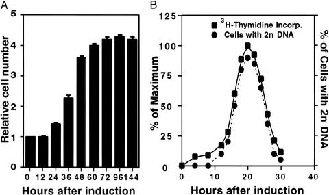
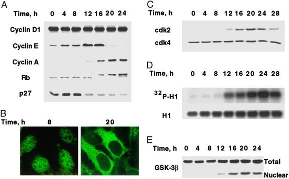
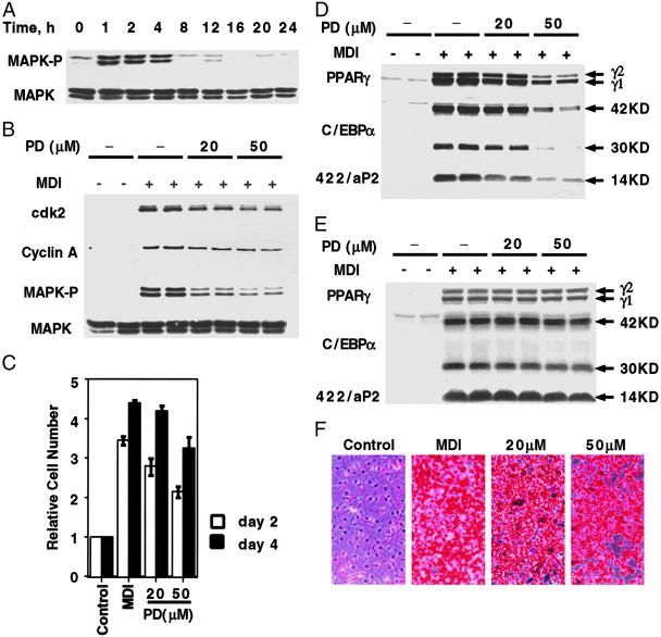
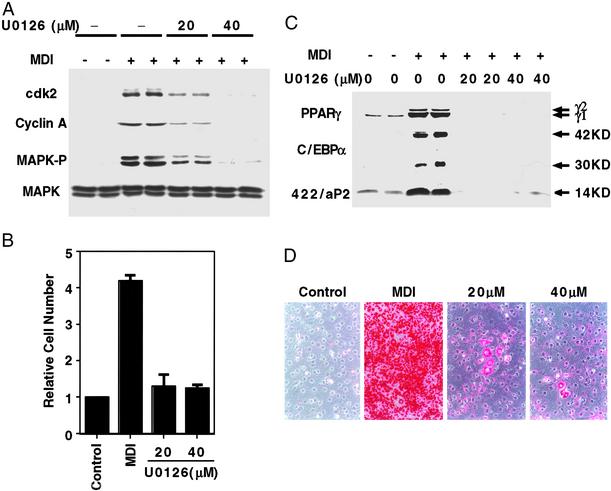
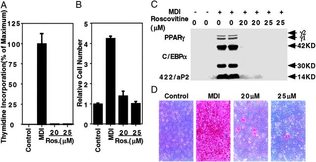
When induced to differentiate, growth-arrested 3T3-L1 preadipocytes synchronously reenter the cell cycle and undergo mitotic clonal expansion (MCE) followed by expression of genes that produce the adipocyte phenotype. The preadipocytes traverse the G(1)S checkpoint synchronously as evidenced by the expressionactivation of cdk2-cyclin-EA, turnover of p27kip1, hyperphosphorylation of Rb, translocation of cyclin D(1) from nuclei to cytoplasm and GSK-3beta from cytoplasm to nuclei, and incorporation of [(3)H]thymidine into DNA. As the cells cross the G(1)S checkpoint, CEBPbeta acquires DNA-binding activity, initiating a cascade of transcriptional activation that culminates in the expression of adipocyte proteins. The mitogen-activated protein kinaseextracellular signal-regulated kinase kinase (MEK) inhibitor PD98059 delays, but does not block, MCE and differentiation, the extent of the delay causing a comparable delay in the expression of cell-cycle markers, MCE, and adipogenesis. The more potent and specific MEK inhibitor UO126 and the cyclin-dependent kinase inhibitor roscovitine, which inhibit the cell cycle at different points, block MCE, expression of cell cycle and adipocyte markers, as well as adipogenesis. These results show that MCE is a prerequisite for differentiation of 3T3-L1 preadipocytes into adipocytes.


Figures

Figure 1
Figure 1
Synchronous reentry of the cell cycle by growth-arrested 3T3-L1 preadipocytes after induction of differentiation. (A) Day 0 postconfluent 3T3-L1 preadipocytes were induced to differentiate into adipocytes by using the standard differentiation protocol. Cell number was determined by using a Coulter counter, and the relative cell number was plotted. At the time of induction, there were 1.4 × 10⁶ cells per 6-cm culture dish. (B) Day 0 postconfluent 3T3-L1 preadipocytes were induced to differentiate. At the times indicated, cells were trypsinized, fixed, and labeled with propidium iodide, and the relative DNA content per cell was determined by FACS analysis. For comparison, FACS analysis data are shown (as % of cells with 2n DNA content) with [3H]thymidine incorporation data from an earlier paper (11).
Figure 2
Figure 2
Changes in the expression and translocation of cell-cycle markers and GSK-3β during MCE. (A) Day 0 postconfluent 3T3-L1 preadipocytes were induced to differentiate into adipocytes. At the times indicated, whole-cell lysates (50 μg of protein) were subjected to SDS/PAGE and immunoblotted. The decreased mobility of Rb after 16 h was shown to be caused by hyperphosphorylation by incubating nuclear extracts with alkaline phosphatase, which increased the mobility of Rb (not shown). (B) 3T3-L1 preadipocytes on glass coverslips were cultured to postconfluence in DMEM containing 10% calf serum and induced to differentiate. At 8 and 20 h, cells were fixed and exposed to cyclin D1 antibody followed by FITC-labeled secondary antibody. (C) At the indicated times after induction of differentiation, cell lysates (50 μg of protein) were subjected to SDS/PAGE immunoblotted with antibodies to cdk2 and cdk4. (D) Nuclear extracts were prepared at the indicated times after induction. Cdk2 was immunoprecipitated and incubated with purified histone H1 and [γ-32P]ATP. After SDS/PAGE, [32P]histone H1 was detected by autoradiography. Total histone H1 was detected by Coomassie blue staining. (E) At the indicated times after induction of differentiation, preadipocytes were lysed and resolved into nuclear and cytoplasmic fractions. The whole-cell lysate and nuclear fractions (identical cell equivalents) were subjected to SDS/PAGE and then immunoblotted with antibody to GSK-3β.
Figure 3
Figure 3
Effect of PD98059, a MEK inhibitor, on MAPK kinase activity, cell cycle markers, MCE, and adipogenesis. (A) Two-day postconfluent 3T3-L1 preadipocytes were induced to differentiate by using the standard differentiation protocol. At various time points, cell lysates were prepared and subjected to SDS/PAGE and immunoblotting with antibodies to MAPK and phospho-MAPK. (B) Two-day postconfluent 3T3-L1 preadipocytes were exposed to 20 or 50 μM PD98059 for 30 min before induction and again at the time of induction. Cell lysates were prepared 1 h later for MAPK and phospho-MAPK and 20 h later for cyclin A and cdk2 immunoblot analyses. (C) Two-day postconfluent 3T3-L1 preadipocytes were exposed to 20 or 50 μM PD98059 for 30 min before induction and again at the time of induction. On days 2 and 4, cell number was determined. MDI refers to the differentiation inducers. (D) Postconfluent 3T3-L1 preadipocytes were treated as in B. Cell lysates were prepared on day 3, and 50 μg of lysate protein were subjected to SDS/PAGE and immunoblotted with antibodies against C/EBPα, PPARγ, and 422/aP2. (E) Cells were treated as in B, above, except that cell lysates were prepared on day 6. (F) Cells were treated as B, except that on day 8, cells were fixed and cytoplasmic triacylglycerol was stained with Oil Red-O. PD98059 concentrations are given (20 and 50 μM).
Figure 4
Figure 4
Effect of U0126 (a MEK inhibitor) on MCE, expression of cell cycle and adipocyte markers, and adipogenesis. (A) Two-day postconfluent 3T3-L1 preadipocytes were incubated with 20 or 40 μM U0126 for 30 min and then induced to differentiate with the same amount of U0126; U0126 was added again 24 h later. Cell lysates were prepared 1 h later for MAPK and phospho-MAPK analysis and at 20 h for cyclin A and cdk2 analysis. Cell lysates (50 μg of protein) were subjected to SDS/PAGE and immunoblotting as described in Fig. 3. (B) Two-day postconfluent 3T3-L1 preadipocytes were treated as A, and cell number was determined on day 4. (C) Two-day postconfluent 3T3-L1 preadipocytes were treated as in A; cell lysates were prepared on day 6 and subjected to analysis as in A with antibodies to C/EBPα, PPARγ, and 422/aP2. (D) Two-day postconfluent 3T3-L1 preadipocytes were treated as in A; on day 8, cells were fixed, and cytoplasmic triacylglycerol was stained with Oil Red-O. U0126 concentrations are given (20 and 40 μM).
Figure 5
Figure 5
Effect of roscovitine (a cyclin-dependent kinase inhibitor) on DNA synthesis, MCE, and adipogenesis. (A) Two-day postconfluent 3T3-L1 preadipocytes were treated with 20 or 25 μM roscovitine at the time of induction of differentiation. After 20 h, cells were pulse-labeled with [3H]thymidine for 30 min and lysed; DNA was precipitated, and 3H incorporation into DNA was determined. (B) Two-day postconfluent 3T3-L1 preadipocytes were treated as in A. Roscovotine was added with medium changes (every other day) until day 4, at which time cell number was determined. (C) Two-day postconfluent 3T3-L1 preadipocytes were treated as in B, and cell lysates were prepared on day 6 and subjected to immunoblotting with antibodies to C/EBPα, PPARγ, and 422/aP2. (D) Two-day postconfluent 3T3-L1 preadipocytes were treated as in C; on day 8, they were fixed, and cytoplasmic triacylglycerol was stained with Oil Red-O. Roscovitine concentrations are given (20 and 25 μM).

References

    1. Shepherd P R, Gnudi L, Tozzo E, Yang H, Leach F, Kahn B B. J Biol Chem. 1993;268:22243–22246. - PubMed
    1. Gnudi L, Shepherd P R, Kahn B B. Proc Nutr Soc. 1996;55:191–199. - PubMed
    1. Bernlohr D A, Bolanowski M A, Kelly T J, Lane M D. J Biol Chem. 1985;260:5563–5567. - PubMed
    1. Cornelius P, MacDougald O A, Lane M D. Annu Rev Nutr. 1994;14:99–129. - PubMed
    1. MacDougald O A, Lane M D. Annu Rev Biochem. 1995;64:345–373. - PubMed

Publication types

MeSH terms