. 2003 Jan 7;100(1):336-41.
doi: 10.1073/pnas.0234600100. Epub 2002 Dec 26.

Chemical nature of nitric oxide storage forms in rat vascular tissue

Affiliations

Chemical nature of nitric oxide storage forms in rat vascular tissue

Juan Rodriguez et al. Proc Natl Acad Sci U S A. .

Abstract

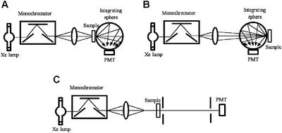
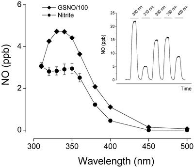
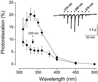
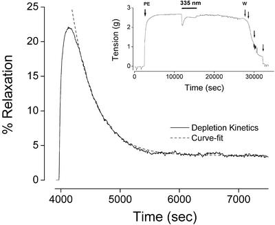
Endothelial NO production results in local formation of adducts that may act as storage forms of NO. Because little is known about their chemical nature, concentrations, and possible role in vascular biology, we sought to characterize those species basally present in rat aorta, using two independent approaches. In the first approach, tissue homogenates were analyzed by using chemiluminescence- and ion-chromatography-based techniques that allow trace-level quantification of NO-related compounds in complex biological matrices. In the second approach, NO stores were characterized by their ability to release NO when illuminated with light and subsequently relax vascular smooth muscle (photorelaxation). The latter included a careful assessment of action spectra for photorelaxation, taking into account the light-scattering properties of the tissue and the storage depletion rates induced by exposure to controlled levels of light. Biochemical analyses revealed that aortic tissues contained 10 +/- 1 microM nitrite, 42 +/- 7 microM nitrate, 40 +/- 6 nM S-nitroso, and 33 +/- 6 nM N-nitroso compounds (n = 4-8). The functional data obtained suggest that the NO photolytically released in the tissue originated from species with photophysical properties similar to those reported for low-molecular-weight S-nitrosothiols, as well as from nitrite. The relative contribution of these potential NO stores to the extent of photorelaxation was consistent with their concentrations detected biochemically in vascular tissue when their photoactivity was taken into account. We conclude that intravascular nitroso species and nitrite both have the potential to release physiologically relevant quantities of NO independent of enzymatic control by NO synthase.


Figures

Figure 1
Figure 1
Diagram of the optical system used for the determination of the optical transmission and the differential pathlength in rat aorta, which were used to compensate photorelaxant responses for internal tissue filtering. Determination of these factors was achieved by measurements of the integrated transmittance (A), diffuse reflectance (B), and collimated transmittance (C) of the sample, and data were processed by using software that simulates the transport of photons via a Monte Carlo technique.
Figure 2
Figure 2
Action spectra for NO release from GSNO and nitrite in 50 mM phosphate buffer, pH 7.4 and 37°C. The photocleaved NO was purged from the reaction vessel with nitrogen and quantified by gas phase chemiluminescence. Data are the means ± SEM of three individual experiments, with error bars being within symbols in the GSNO spectrum. (Inset) Typical tracing obtained with 10 μM GSNO and brief exposures to light of varying wavelengths.
Figure 3
Figure 3
(Inset) Typical “photorelaxation fingerprint” responses obtained in precontracted rat aortic rings exposed to brief challenges with light of varying wavelengths. The average action spectra derived from such tracings are shown, following corrections for the wavelength dependence of the intensity of the light source, the transmission of the organ bath, and the internal filtering within the tissue. The percent photorelaxation refers to percent change from contractile tone before illumination. The upper curve represents the average spectrum of 67 rings from 23 animals obtained with 30-s exposures at each wavelength (means ± SEM). The lower curve represents the average action spectrum of 12 rings from seven animals obtained after 60-min exposure to light at 335 nm (0.3 mW/cm2 irradiance).
Figure 4
Figure 4
(Inset) Representative tracing of a precontracted rat aortic segment that has been exposed for 60 min to 335-nm light at an irradiance of 0.3 mW/cm2 (PE, phenylephrine; W, washout). The depletion kinetics of the photorelaxant response observed during the continuous light exposure is magnified and represented as percent relaxation in the main panel (solid curve). The dashed curve represents a curve-fit to a theoretical optical depletion model (Eq. 4).

Similar articles

Cited by

References

    1. Stamler J S, Jaraki O, Osborne J, Simon D I, Keaney J, Vita J, Singel D, Valeri C R, Loscalzo J. Proc Natl Acad Sci USA. 1992;89:7674–7677. - PMC - PubMed
    1. Minamiyama Y, Takemura S, Inoue M. Biochem Biophys Res Commun. 1996;225:112–115. - PubMed
    1. Jia L, Bonaventura C, Bonaventura J, Stamler J S. Nature. 1996;380:221–226. - PubMed
    1. Furchgott R F, Sleaton W, Jr, McCaman M W, Elchleff J. J Pharmacol Exp Ther. 1955;113:22–23.
    1. Furchgott R F, Ehrreich S J, Grennblatt E. J Gen Physiol. 1961;44:499–519. - PMC - PubMed

Publication types

LinkOut - more resources