. 2003 Jan;77(2):915-22.
doi: 10.1128/jvi.77.2.915-922.2003.

Adenovirus type 5 DNA binding protein stimulates binding of DNA polymerase to the replication origin

Affiliations

Adenovirus type 5 DNA binding protein stimulates binding of DNA polymerase to the replication origin

Bas van Breukelen et al. J Virol. 2003 Jan.

Abstract

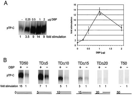
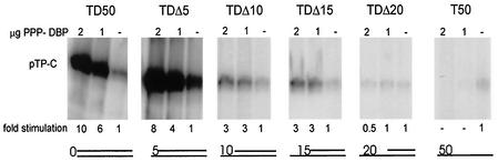
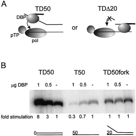
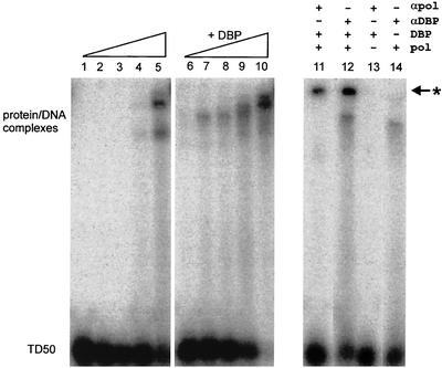
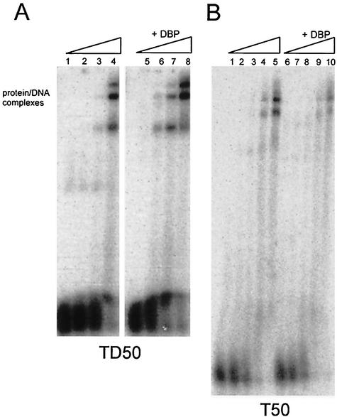
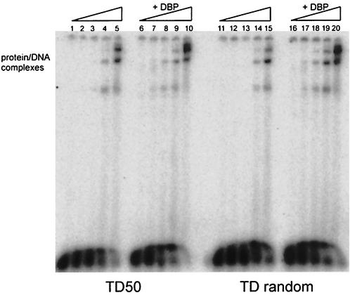
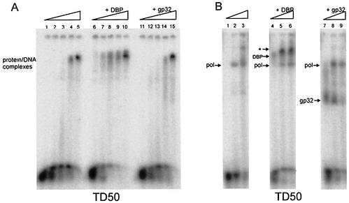
The adenovirus (Ad) DNA-binding protein (DBP) is essential for the elongation phase of Ad DNA replication by unwinding the template in an ATP-independent fashion, employing its capacity to form multimers. DBP also enhances the rate of initiation, with the highest levels obtained at low concentrations of Ad DNA polymerase (Pol). Here, we show that stimulation of initiation depends on the template conformation. Maximal stimulation, up to 15-fold, is observed on double-stranded or viral TP-containing origins. The stimulation is reduced on partially single-stranded origins and DBP does not enhance initiation any more once the origin is completely unwound. This suggests a role for DBP in origin unwinding that is comparable to its unwinding capacity during elongation. However, mutant DBP proteins defective in unwinding and elongation can still enhance initiation on ds templates. DBP also stimulates the binding of nuclear factor I (NFI) to the origin and lowers the K(m) for coupling of the first nucleotide to the precursor terminal protein by Pol. Mobility shift experiments reveal that DBP stimulates the binding of Pol on double-stranded origin and nonorigin DNA but not on single-stranded DNA. This effect is specific for DBP and is also seen with other DNA Pols. Our results suggest that, rather than by origin unwinding, DBP enhances initiation by modulating the origin conformation such that DNA Pol can bind more efficiently.


Figures

FIG. 1.
FIG. 1.
(A) Initiation assay on 0.3 pmol of the TD50 template with various concentrations of DBP. A representative initiation assay is shown. The average level of initiation based on three independent experiments is graphically represented. (B) Initiation assay on origin-containing templates with increasing 5′ gaps. Equimolar amounts of DNA templates (0.3 pmol) were used. The fold stimulation of initiation by DBP of a representative experiment is shown. The pTP-C signal without the addition of DBP was set to 1 for each template. The basal level of initiation (no DBP) on TD50 was set to 1, and the basal levels of initiation of the other templates were determined as follows: TDΔ5, 2.5; TDΔ10, 0.6; TDΔ15, 0.7; TDΔ20, 0.3; and T50, 0.3.
FIG. 2.
FIG. 2.
Initiation assay with the unwinding defective PPP-DBP mutant on 0.3 pmol of origin-containing templates. Indicated are the 5′ gaps ranging from 0 to 50 nucleotides. The amount of stimulation of initiation is shown. The pTP-C signal without the addition of PPP-DBP was set to 1 for each template. The basal level of initiation (no PPP-DBP) on TD50 was set to 1, and the basal levels of initiation of the other templates were determined as follows: TDΔ5, 3.3; TDΔ10, 0.4; TDΔ15, 0.3; TDΔ20, 0.3; and T50, 0.4.
FIG. 3.
FIG. 3.
(A) Model for transient DBP-Pol interaction via the displaced strand. On the left, DBP is able to contact Pol via the displaced strand and thereby stimulates the initiation. On the right, DBP is unable to contact Pol when the ssDNA gap becomes too large, such as in TDΔ20; hence, no stimulation of initiation can be observed. (B) Initiation assay on 0.3 pmol of origin-containing templates. Assay conditions are as described in the legends to Fig. 1 and 2. The fold stimulation of initiation by DBP is shown.
FIG. 4.
FIG. 4.
EMSA with TD50 and increasing Pol concentrations (0, 25, 50, 100, and 200 ng). Lanes 1 to 5 contain no DBP and show DNA-Pol complexes only. The reactions in lanes 6 to 10 contain an additional 20 ng of DBP. Supershifted complexes with antibodies to Pol (lane 11) or DBP (lane 12) are indicated with an asterisk. The “+” symbol indicates the addition of DBP (20 ng), Pol (50 ng), or antibodies.
FIG. 5.
FIG. 5.
(A) EMSA with TD50 and increasing Pol (exo mutant) concentrations (0, 25, 50, and 100 ng). Lanes 1 to 4 contain no DBP and show DNA-Pol complexes only. The reactions in lanes 5 to 8 contain an additional 15 ng of DBP. (B) EMSA with increasing Pol (exo mutant) concentrations (0, 25, 50, 100, or 200 ng) preincubated with T50 ssDNA either without DBP (lanes 1 to 5) or with 0.6 ng of DBP (lanes 6 to 10). Note that the DBP concentration is lower in EMSAs on ssDNA because the affinity of DBP for ssDNA is much higher than that for dsDNA.
FIG. 6.
FIG. 6.
EMSA with increasing Pol concentrations (0, 12.5, 25, 50, or 100 ng) preincubated either with TD50 or a 50-bp random dsDNA probe (TD random), without DBP (lanes 1 to 5 and 11 to 15) or with 15 ng of DBP (lanes 6 to 10 and 16 to 20).
FIG. 7.
FIG. 7.
(A) EMSA of the template TD50, preincubated with increasing phage T4 Pol concentrations (0, 3.5, 7, 14, or 28 ng) without SSB (lanes 1 to 5), in the presence of 15 ng of DBP (lanes 6 to 10), or in the presence of 40 ng of phage T4 gp32 (lanes 11 to 15). (B) EMSA of the template TD50 preincubated with increasing Ad5 Pol concentrations (0, 50, and 100 ng) without SSB (lanes 1 to 3) or in the presence of 15 ng of DBP (lanes 4 to 6) or 40 ng of phage T4 gp32. Arrows indicate the single-protein DNA complexes. Only in lanes 5 and 6 is a complex consisting of DNA, DBP and Pol present (✽).

Similar articles

Cited by

References

    1. Anderson, C. W., M. M. Hardy, J. J. Dunn, and D. F. Klessig. 1983. Independent, spontaneous mutants of adenovirus type 2-simian virus 40 hybrid Ad2+ND3 that grow efficiently in monkey cells possess identical mutations in the adenovirus type 2 DNA-binding protein gene. J. Virol. 48:31-39. - PMC - PubMed
    1. Ariga, H., H. Klein, A. J. Levine, and M. S. Horwitz. 1980. A cleavage product of the adenovirus DNA binding protein is active in DNA replication in vitro. Virology 101:307-310. - PubMed
    1. Armentero, M. T., M. Horwitz, and N. Mermod. 1994. Targeting of DNA polymerase to the adenovirus origin of DNA replication by interaction with nuclear factor I. Proc. Natl. Acad. Sci. USA 91:11537-11541. - PMC - PubMed
    1. Bosher, J., I. R. Leith, S. M. Temperley, M. Wells, and R. T. Hay. 1991. The DNA-binding domain of nuclear factor I is sufficient to cooperate with the adenovirus type 2 DNA-binding protein in viral DNA replication. J. Gen. Virol. 72:2975-2980. - PubMed
    1. Brenkman, A. B., E. C. Breure, and P. C. van der Vliet. 2002. Molecular architecture of adenovirus DNA polymerase and location of the protein primer. J. Virol. 76:8200-8207. - PMC - PubMed

Publication types

LinkOut - more resources