. 2003 Jan;77(2):1578-83.
doi: 10.1128/jvi.77.2.1578-1583.2003.

Hepatitis C virus f protein is a short-lived protein associated with the endoplasmic reticulum

Affiliations

Hepatitis C virus f protein is a short-lived protein associated with the endoplasmic reticulum

Zhenming Xu et al. J Virol. 2003 Jan.

Abstract

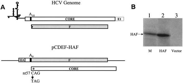
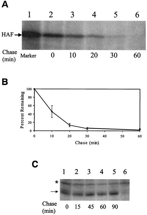
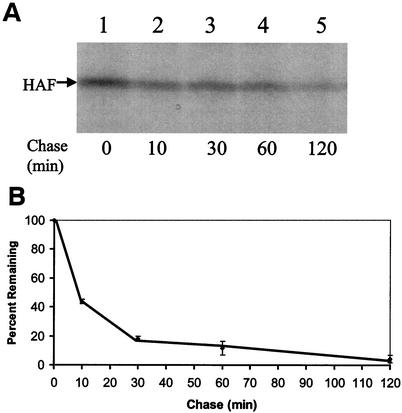
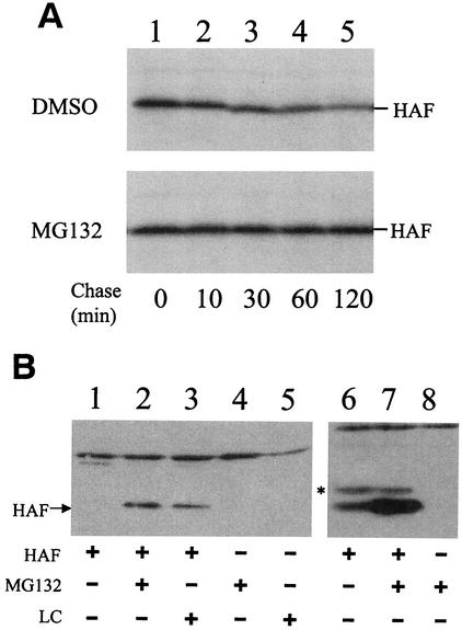
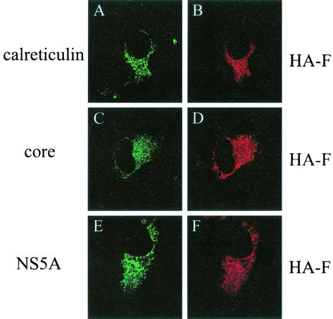
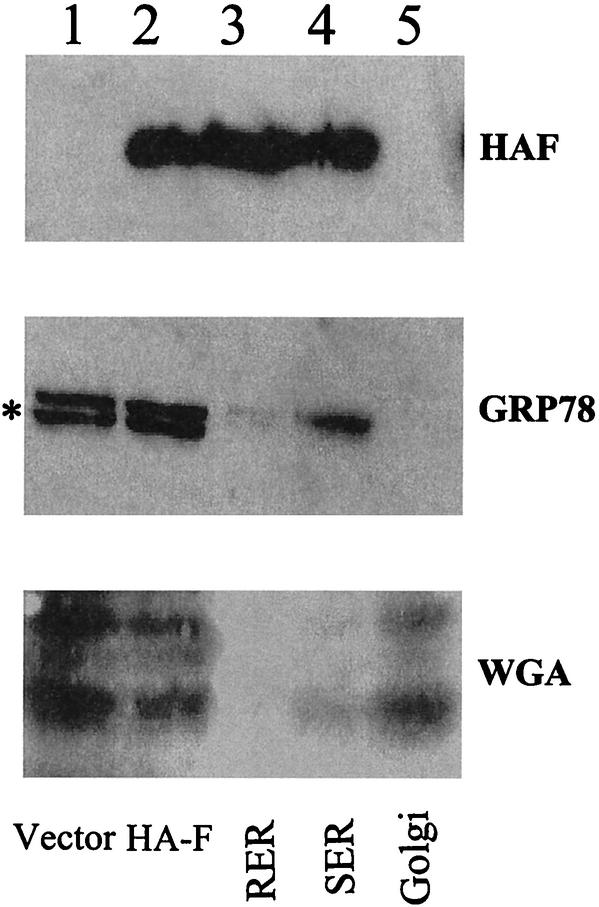
Hepatitis C virus (HCV) F protein is a newly discovered HCV gene product that is expressed by translational ribosomal frameshift. Little is known about the biological properties of this protein. By performing pulse-chase labeling experiments, we demonstrate here that the F protein is a labile protein with a half-life of <10 min in Huh7 hepatoma cells and in vitro. The half-life of the F protein could be substantially increased by proteasome inhibitors, suggesting that the rapid degradation of the F protein is mediated by the proteasome pathway. Further immunofluorescence staining and subcellular fractionation experiments indicate that the F protein is primarily associated with the endoplasmic reticulum. This subcellular localization is similar to those of HCV core and NS5A proteins, raising the possibility that the F protein may participate in HCV morphogenesis or replication.


Figures

FIG. 1.
FIG. 1.
Expression of the HA-F protein in Huh7 hepatoma cells. (A) Illustrations of the 5′ end of the HCV-1 genome (top) and the HA-F cDNA construct (bottom). The HCV genome shown contains the 5′ noncoding region and the coding regions of the core protein and the F protein. The F protein coding sequence is shaded. A10, the stretch of 10 adenosines at codons 8 to 11 of the core protein coding sequence. This sequence contains the ribosomal frameshift signal for the synthesis of the F protein (28). The HCV-1 genome was used for the construction of the plasmid pCDEF-HAF. In this construct, one adenosine was deleted from A10 to generate A9. This deletion fused the first 10 codons of the core protein to the F protein coding sequence. The HA tag, indicated by a stippled box, was fused to the 5′ end of the coding sequence. The location of the nt 57 C-to-T mutation is indicated by an asterisk. This mutation created a TAG termination codon in the core protein sequence. (B) Immunoprecipitation of the HA-F protein. pCDEF-HAF (lane 2) or the control vector pCDEF (lane 3) was transfected into Huh7 cells by using CaPO4 precipitation procedures (13). Cells were starved for methionine for 3 h at 2 days after transfection and then radiolabeled with [35S]methionine for 1 h, followed by immunoprecipitation with anti-HA antibody (13). The [35S]methionine-labeled HA-F protein synthesized in vitro with rabbit reticulocyte lysates (28) was shown in lane 1 to serve as a marker (M).
FIG. 2.
FIG. 2.
Pulse-chase labeling experiment of the HCV proteins expressed in Huh7 cells. (A) Pulse-chase labeling experiment of the HA-F protein. Huh7 cells transfected with pCDEF-HAF by CaPO4 precipitation were pulse-labeled with [35S]methionine for 10 min and chased with unlabeled methionine for 0, 10, 20, 30, and 60 min (lanes 2 to 6). Cells were then lysed for immunoprecipitation with the anti-HA antibody by our previous procedures (14). Lane 1 is the [35S]methionine-labeled HA-F protein marker, which was synthesized in vitro by using the rabbit reticulocyte lysates. (B) Determination of the half-life of the HA-F protein. The autoradiogram shown in panel A was analyzed with SigmaScan. The results represented the average of three independent experiments. The HA-F protein level at the zero time point of chase was defined as 100%. (C) Pulse-chase labeling experiment of the HA-core protein. Huh7 cells transfected with pCDEF-HA-core (13) were pulse-labeled with [35S]methionine for 10 min and chased with unlabeled methionine for 0, 15, 45, 60, and 90 min (lanes 1 to 5). Lane 6 was Huh7 cells transfected with the control pCDEF vector. The asterisk marks the location of a nonspecific protein band. The arrow denotes the core protein band.
FIG. 3.
FIG. 3.
Pulse-chase labeling experiments of the HA-F protein synthesized in vitro. (A) Autoradiograms of the pulse-chase experiments. The HA-F coding sequence was inserted into pRc/CMV (Invitrogen). The HA-F RNA was then synthesized by using the T7 RNA polymerase and translated with the rabbit reticulocyte lysates. Details of these experimental procedures had been described (28). The HA-F protein was pulse-labeled with [35S]methionine for 10 min. The translation reaction was then stopped by the addition of cycloheximide to a final concentration of 400 μM. The HA-F protein was then chased for 0, 10, 30, 60, or 120 min (lanes 1 to 5). (B) Half-life of the HA-F protein in vitro. The results shown in panel A were quantified with SigmaScan. The results represented the average of three independent experiments. The HA-F protein level at the zero time point was defined as 100%.
FIG. 4.
FIG. 4.
Stabilization of the HA-F protein by proteasome inhibitors. (A) Pulse-chase labeling experiments of HA-F synthesized in vitro. The HA-F protein was pulse-labeled with [35S]methionine for 10 min as described in the legend to Fig. 3. The translation reactions were then stopped with cycloheximide. MG132 (lower panel) or its control solvent DMSO (upper panel) was then added to a final concentration of 100 μg/ml. The reaction mixtures were chased for 0, 10, 30, 60, or 120 min (lanes 1 to 5). (B) Stabilization of HA-F by proteasome inhibitors in Huh7 cells. pCDEF-HAF (lanes 1 to 3), its control vector pCDEF (lanes 4, 5, and 8), or pCDEF-9aCore (lanes 6 and 7) was transfected into Huh7 cells. pCDEF-9aCore is identical to pCDEF-HAF with the exception that it does not have the C-to-T mutation at nt 57 (28). At 48 h after transfection, cells were treated with DMSO (lane 1), 1 μg of MG132/ml (lanes 2 and 4), or 20 μM of lactacystin (LC) (lanes 3 and 5) for 6 h. Cells were then lysed for Western blot analysis with the anti-HA antibody by using our previous procedures (13). The protein signals were analyzed by the enhanced chemiluminescence kit (Pierce). The expression level of HAF was low in DMSO-treated cells. Hence, lanes 6 to 8 were purposely overexposed on the film to reveal the HAF protein in DMSO-treated cells (lane 6). The asterisk marks the location of the HA-core protein. The identity of this protein was confirmed by Western blotting with the anti-core antibody (data not shown).
FIG. 5.
FIG. 5.
Immunofluorescence double-staining analysis for the subcellular localization of the HA-F protein. (A and B) Huh7 cells transfected with pCDEF-HAF were double stained with rabbit anti-calreticulin and mouse anti-HA primary antibodies and fluorescein isothiocyanate (FITC)-conjugated goat anti-rabbit and rhodamine (RITC)-conjugated goat anti-mouse secondary antibodies. (C and D) Huh7 cells cotransfected with pCDEF-HAF and pCDEF-core (13) were double stained with rabbit anti-HCV core and mouse anti-HA primary antibodies and FITC-conjugated goat anti-rabbit and RITC-conjugated goat anti-mouse secondary antibodies. (E and F) Huh7 cells containing the HCV subgenomic RNA replicon (unpublished data) were transfected with pCDEF-HAF and double stained with mouse anti-NS5A and rat anti-HA primary antibodies and FITC-conjugated goat anti-mouse and RITC-conjugated goat anti-rat secondary antibodies. In all cases, cells were fixed in 3.7% formaldehyde in phosphate-buffered saline (PBS) at 48 h after transfection for staining by using our previous procedures (14). The HA-F protein was stained in red, whereas all of the other proteins were stained in green. The images were captured with a Nikon confocal microscope at the USC Liver Center.
FIG. 6.
FIG. 6.
Membrane fractionation experiments for the analysis of the subcellular localization of HA-F. Huh7 cells transfected with pCDEF-HAF were rinsed with PBS and scraped off the plates into PBS. After a brief centrifugation at 1,500 × g, cells were homogenized with a Dounce homogenizer in a 0.25 M sucrose solution containing 10 mM HEPES (pH 7.4), 1 mM EDTA, and 1 mM phenylmethylsulfonyl fluoride. After a brief centrifugation at 15,000 × g, the postnuclear supernatant was loaded on a discontinuous sucrose gradient containing 0.6, 1.0, 1.3, and 2.0 M sucrose in 10 mM HEPES (pH 7.4). The gradient was centrifuged at 40,000 rpm by using a Beckman SW40 Ti rotor for 2 h at 4°C as previously described (26). Lane 1, cells transfected with the control vector pCDEF; lanes 2 to 5, cells transfected with pCDEF-HAF; lanes 1 and 2, the postnuclear supernatant prior to fractionation; lane 3, the rough ER (RER) fraction isolated from the 1.3-2.0 M sucrose interface; lane 4, the smooth ER (SER) fraction isolated from the 1.0-1.3 M sucrose interface; lane 5, the Golgi fraction isolated from the 0.6-1.0 M sucrose interface. (Top panel) Western blot analysis with anti-HA antibody; (middle panel) Western blot analysis with anti-GRP78 antibody; (bottom panel) Western blot analysis with horseradish peroxidase-conjugated wheat germ agglutinin (WGA). The asterisk denotes a nonspecific protein band. This protein band did not cofractionate with GRP78 into the ER membrane fractions. Wheat germ agglutinin reacted with multiple protein bands.
FIG. 7.
FIG. 7.
Hydrophilicity plot of the F protein. The HCV-1 F protein coding sequence with 1 nt deletion in the 10-A stretch was analyzed by the MacVector program. The two thick lines highlight the two major hydrophobic domains.

Similar articles

Cited by

References

    1. Blight, K. J., A. A. Kolykhalov, and C. M. Rice. 2000. Efficient initiation of HCV RNA replication in cell culture. Science 290:1972-1974. - PubMed
    1. Blight, K. J., A. Grakoui, H. L. Hanson, and C. M. Rice. 2002. The molecular biology of hepatitis C virus, p. 81-108. In J.-H. J. Ou (ed.), Hepatitis viruses. Kluwer Academic Publishers, Norwell, Mass.
    1. Brass, V., E. Bieck, R. Montserret, B. Wolk, J. A. Hellings, H. E. Blum, F. Penin, and D. Moradpour. 2002. An amino-terminal amphipathic alpha-helix mediates membrane association of the hepatitis C virus nonstructural protein 5A. J. Biol. Chem. 277:8130-8139. - PubMed
    1. Carrere-Kremer, S., C. Montpellier-Pala, L. Cocquerel, C. Wychowski, F. Penin, and J. Dubuisson. 2002. Subcellular localization and topology of the p7 polypeptide of hepatitis C virus. J. Virol. 76:3720-3730. - PMC - PubMed
    1. Cocquerel, L., A. Op de Beeck, M. Lambot, J. Roussel, D. Delgrange, A. Pillez, C. Wychowski, F. Penin, and J. Dubuisson. 2002. Topological changes in the transmembrane domains of hepatitis C virus envelope glycoproteins. EMBO J. 21:2893-2902. - PMC - PubMed

Publication types

LinkOut - more resources