. 2003 Jan;77(2):1589-94.
doi: 10.1128/jvi.77.2.1589-1594.2003.

Proline 78 is crucial for human immunodeficiency virus type 1 Nef to down-regulate class I human leukocyte antigen

Affiliations

Proline 78 is crucial for human immunodeficiency virus type 1 Nef to down-regulate class I human leukocyte antigen

Takeshi Yamada et al. J Virol. 2003 Jan.

Abstract

Human immunodeficiency virus type 1 Nef down-regulates human leukocyte antigen class I (HLA-I) in T lymphocytes, and the down-regulation involves the Nef proline-rich domain (PRD) containing four prolines at positions 69, 72, 75, and 78. We used a Sendai virus vector with nef and examined regulation by Nef of HLA-I and CD4 in suspension cultures of cells such as T lymphocytes. Analyses of a series of PRD substitution mutants indicated that, because the substitution of Pro78 with Ala abolished down-regulation of HLA-I but not of CD4, Pro78 is important for HLA-I down-regulation in T lymphocytes.


Figures
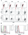
FIG. 1.
FIG. 1.
Flow-cytometric analysis of CD4 and HLA-I expression on cell surfaces. CEM cells were infected with each rSeV at a multiplicity of infection of 10 for 1 h. Twenty-four hours after infection, cells were treated at 4°C for 20 min with an allophycocyanin-labeled anti-CD4 antibody (Becton Dickinson Immunocytometry Systems, San Jose, Calif.), and an R-phycoerythrin-labeled anti-HLA-I antibody (W6/32; Dako, Glostrup, Denmark) against a monomorphic epitope of HLA-A, -B, and -C or an anti-HLA-C antibody (29) with an R-phycoerythrin-labeled secondary antibody (Dako). A FACSCalibur flow cytometer (Becton Dickinson Immunocytometry Systems) with CellQuest software (Becton Dickinson Immunocytometry Systems) was used for flow cytometry. Isotype-matched control antibodies were included to detect nonspecific binding to cells. FlowJo software (Tree Star, San Carlos, Calif.) was used to make configurations. (A) Cells were analyzed for fluorescence intensity in the cell population gated as shown by a circle in each upper panel. The vertical axis represents signal intensity obtained with anti-HLA-I or anti-HLA-C. The horizontal axis represents signal intensity obtained with an anti-CD4 antibody. (B) Histograms of signal intensity obtained with anti-HLA-I or anti-HLA-C. Histograms are overlaid with a dotted-line histogram of an immunoglobulin isotype control. Values are mean fluorescence intensities (MFI) of HLA-I and HLA-C on the cell surface. The data were reproduced in three independent experiments. mock, SeV without inserted genes; Nef, rSeV with wild-type nef gene.
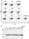
FIG. 2.
FIG. 2.
Analysis of HLA-I down-regulation in CEM cells infected with a series of rSeVs carrying mutant nef. CEM cells were infected with each rSeV at a multiplicity of infection of 10 for 1 h. (A) Flow-cytometric analysis of CD4 and HLA-I expression on the cell surface 24 h after infection. The vertical axis represents signal fluorescent intensity obtained with an anti-HLA-I antibody. The horizontal axis represents signal intensity obtained with an anti-CD4 antibody. (B) Cellular expression of Nef proteins by infection with rSeVs. CEM cells (106) were infected with each rSeV at a multiplicity of infection of 10. One-tenth of the cell lysates was separated by electrophoresis in a sodium dodecyl sulfate-10% polyacrylamide gel. The separated proteins were blotted to a nitrocellulose membrane and stained with an anti-Nef antibody. The arrow indicates bands of about 27 kDa, corresponding to the Nefs shown at the top. The positions of size markers are shown on the left.
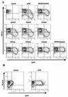
FIG. 3.
FIG. 3.
Flow-cytometric analysis of HIV-1-infected CEM-GFP cells. (A) The vertical axis represents signal intensity obtained with an anti-HLA-I. The horizontal axis represents signal intensity obtained with GFP fluorescence; GFP expression is achieved by HIV-1 infection. The wild-type and mutant Nefs were expressed in the context of HIV-1NL432. (B) Surface levels of HLA-C of the cells infected with the wild-type HIV-1NL432 were assayed. The vertical axis represents signal intensity obtained with an anti-HLA-C antibody. The horizontal axis represents signal intensity of GFP fluorescence. The results are representative of three independent experiments. mock, non-HIV-1-infected CEM-GFP cells.

References

    1. Adachi, A., H. E. Gendelman, S. Koenig, T. Folks, R. Willey, A. Rabson, and M. A. Martin. 1986. Production of acquired immunodeficiency syndrome-associated retrovirus in human and nonhuman cells transfected with an infectious molecular clone. J. Virol. 59:284-291. - PMC - PubMed
    1. Aiken, C., J. Konner, N. R. Landau, M. E. Lenburg, and D. Trono. 1994. Nef induces CD4 endocytosis: requirement for a critical dileucine motif in the membrane-proximal CD4 cytoplasmic domain. Cell 76:853-864. - PubMed
    1. Cesarman, E., Y. Chang, P. S. Moore, J. W. Said, and D. M. Knowles. 1995. Kaposi's sarcoma-associated herpesvirus-like DNA sequences in AIDS-related body-cavity-based lymphomas. N. Engl. J. Med. 332:1186-1191. - PubMed
    1. Collins, K. L., and D. Baltimore. 1999. HIV's evasion of the cellular immune response. Immunol. Rev. 168:65-74. - PubMed
    1. Collins, K. L., B. K. Chen, S. A. Kalams, B. D. Walker, and D. Baltimore. 1998. HIV-1 Nef protein protects infected primary cells against killing by cytotoxic T lymphocytes. Nature 391:397-401. - PubMed

Publication types

MeSH terms

Substances