. 2003 Jan 2;22(1):13-23.
doi: 10.1093/emboj/cdg007.

Control of protein life-span by N-terminal methionine excision

Affiliations

Control of protein life-span by N-terminal methionine excision

Carmela Giglione et al. EMBO J. .

Abstract

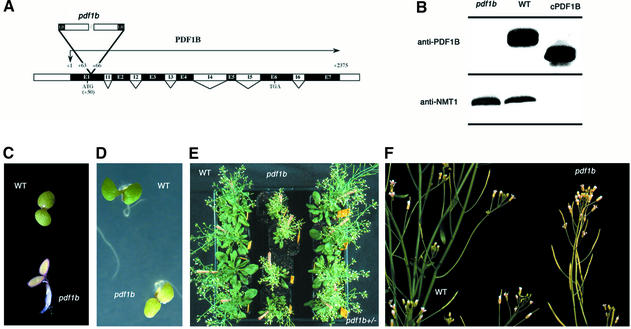
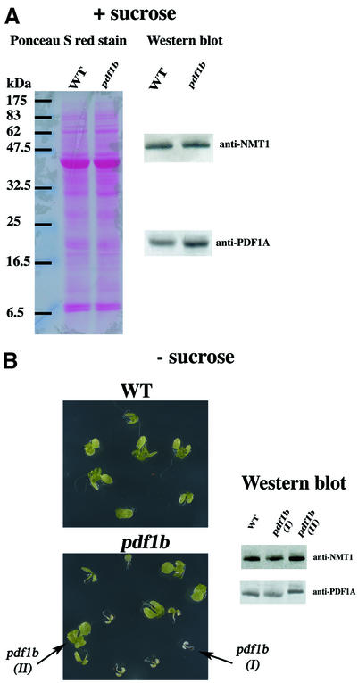
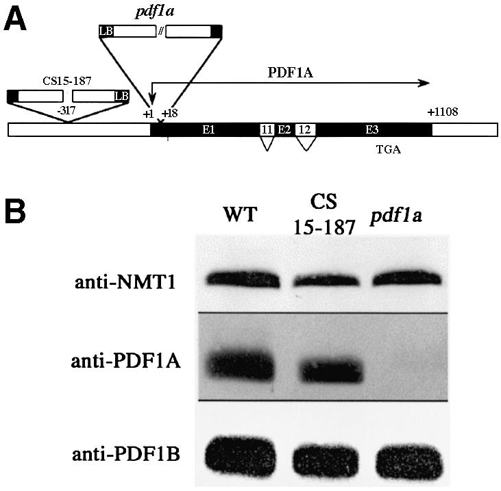
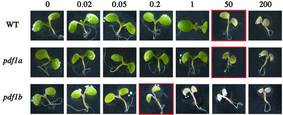
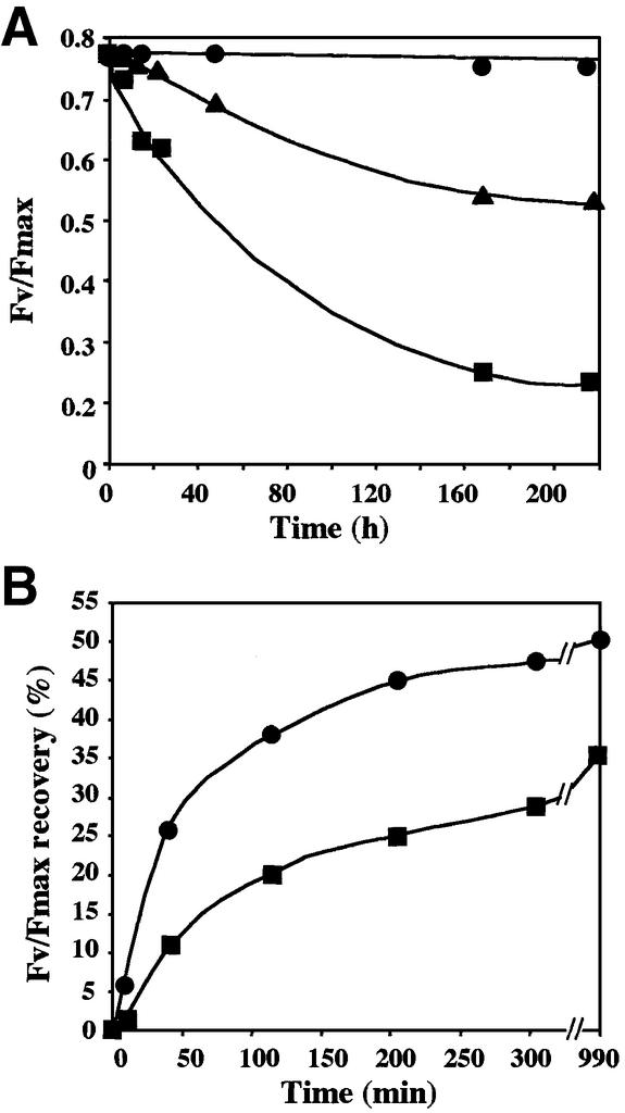
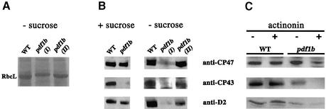
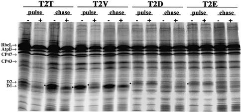
Peptide deformylases (PDFs) have been discovered recently in eukaryotic genomes, and it appears that N-terminal methionine excision (NME) is a conserved pathway in all compartments where protein synthesis occurs. This work aimed at uncovering the function(s) of NME in a whole proteome, using the chloroplast-encoded proteins of both Arabidopsis thaliana and Chlamydomonas reinhardtii as model systems. Disruption of PDF1B in A.thaliana led to an albino phenotype, and an extreme sensitivity to the PDF- specific inhibitor actinonin. In contrast, a knockout line for PDF1A exhibited no apparent phenotype. Photosystem II activity in C.reinhardtii cells was substantially reduced by the presence of actinonin. Pulse-chase experiments revealed that PDF inhibition leads to destabilization of a crucial subset of chloroplast-encoded photosystem II components in C. reinhardtii. The same proteins were destabilized in pdf1b. Site-directed substitutions altering NME of the most sensitive target, subunit D2, resulted in similar effects. Thus, plastid NME is a critical mechanism specifically influencing the life-span of photosystem II polypeptides. A general role of NME in modulating the half-life of key subsets of proteins is suggested.


Figures

None
Fig. 1. Characterization of the A.thaliana pdf1b line. (A) Schematic representation of pdf1b gene disruption in A.thaliana line pdf1b. The exon–intron structure is shown. Translation initiation and termination codons are indicated. The T-DNA inserts are represented with the 5′ border sequences of the insert (LB) labeled to indicate the orientation of the insertion. A three-base-pair deletion was observed at the site of the insertion as indicated. (B) Presence of PDF1B in wild-type and line pdf1b. Arabidopsis seeds were synchronized in the dark at 4°C for 2 days before sowing. Four hundred milligrams of 2-week-old shoots were homogenized and total proteins were extracted as described. Aliquots (250 µg) of protein were analyzed by SDS–PAGE; 250 ng of cPDF1B, the purified catalytic domain of PDF1B (Serero et al., 2001a), was run in parallel. The gels were blotted and analyzed by western blotting with anti-PDF1B and anti-NMT1 as a control. (C) Albino phenotype of 2-day-old pdf1b plantlet compared with wild type. (D) Intermediate phenotype of 3-day-old pdf1b plantlet compared with wild type. (E) Forty-five-day-old wild-type, pdf1b+/– and pdf1b plantlets, seen from the top. (F) Forty-five-day-old wild-type and pdf1b plantlets, seen from the side.
None
Fig. 2. KO pdf1b plants counteract the absence of PDF1B by increasing the level of PDF1A. (A) Expression of PDF1A in wild-type and pdf1b plants grown in 1% sucrose medium. One hundred and fifty micrograms of total protein was separated by SDS–PAGE, transferred to a nitrocellulose membrane and stained with Ponceau S red stain. The membrane was destained and probed using anti-PDF1A and anti-NMT1 antibodies. (B) Wild-type and pdf1b seeds were sown in a sucrose-minus growth medium, synchronized in the dark at 4°C for 2 days and then incubated in a growth cabinet. Seedlings were photographed 2 weeks later and each phenotype was collected separately (I, albino; II, greening). Aliquots (150 µg) of total protein extract were analyzed by 14% SDS–PAGE and western blotting as in (A).
None
Fig. 3. Characterization of the A.thaliana pdf1a line. (A) Schematic representation of PDF1A gene disruption in A.thaliana lines CS1813– 43 and CS15–187. The exon–intron structure is shown as in Figure 1. Translation initiation and termination codons are indicated. The T-DNA inserts are represented with the 5′ border sequences of the insert (LB) labeled to indicate the orientation. (B) PDF1A protein in A.thaliana wild-type, pdf1a and CS15–187 lines. Aliquots (250 µg) of total protein extracts were analyzed by 14% SDS–PAGE. Western blots were performed with anti-PDF1A, anti-PDF1B and anti-NMT1 antibodies.
None
Fig. 4. Line pdf1b is hypersensitive to actinonin. Plants were grown for 5 days in the presence of the indicated (top) concentration of actinonin (µM): (A) wild type; (Bpdf1a line; (Cpdf1b line.
None
Fig. 5. Actinonin leads to a reduction of PSII efficiency in C.reinhardtii. (A) Measurement of the variable fluorescence parameter Fv/Fmax upon actinonin addition (circles, 0 µM; triangles, 50 µM; squares, 500 µM) at time zero in early exponential-phase cultures. (B) Photo-inhibition experiments were carried out for 30 min when the variable fluorescence reached zero. The time-course of recovery in the absence (circles) or the presence (squares) of 0.5 mM actinonin was followed. To visualize only the fraction of recovery that depends on de novo protein synthesis (70%), we subtracted the Fv/Fmax measured at each time point in a duplicate sample incubated with 20 µg/ml chloramphenicol.
None
Fig. 6. Inhibition of PDF induces a rapid degradation of plastid-encoded PSII subunits in C.reinhardtii. The experiments were performed in the presence (+) or the absence (–) of 0.5 mM actinonin. The location of several plastid proteins identified by western blot is indicated. (A) Samples were 45 min pulse-labeled with [14C]acetate 48 h after addition of actinonin. A phosphoimage of the urea-SDS–PAGE (12%–18% acrylamide) is shown. (B) Time-course pulse-labeling with [14C]acetate was performed 10 min after addition of actinonin. The contrast in the lower part of the gel was intensified to show PsbH and its derivatives, PsbH′ and PsbH”, more clearly. (C) Quantification of the data reported in (B) for CP43, CP47, D1, D2 (D2.1 + D2.2), RbcL and AtpB. (D) Pulse–chase labeling of whole cells with [35S]sulfate. A close-up of the phosphoimage of the SDS–PAGE (7.5%–15% acrylamide) in the region of the RbcL band is shown. AtpA and AtpB are not separated under these conditions and co-migrate just below RbcL. (E) Phosphoimage of [35S]sulfate-labeled membrane fractions analyzed by two-dimensional PAGE involving native (horizontal; top right) followed by denaturing conditions of separation (vertical). D1 and D2 were localized on the gels by western blotting analysis with specific antibodies (data not shown).
None
Fig. 7. The accumulation of PSII components is affected in A.thaliana pdf1b lines as in actinonin-treated C.reinhardtii cells. Plants were grown for 15 days. Proteins were analyzed by SDS–PAGE. Western blot analysis was performed using anti-D2, anti-CP43 and anti-CP47 antibodies. (A) Wild-type and pdf1b lines were grown in the absence of sucrose. Plantlets were collected as described in Figure 2. Aliquots (150 µg) of total proteins were loaded on the gel, transferred onto a nitrocellulose membrane and stained with Ponceau S red stain. The position of RbcL is indicated. (B) The destained nitrocellulose membrane from (A) was analyzed by western blotting. (C) Wild-type and pdf1b lines were grown in the absence (–) or the presence (+) of 1 µM actinonin. One-hundred-milligram samples of membrane proteins were analyzed by western blotting.
None
Fig. 8. Inhibiting D2 N-terminal methionine excision by changing its second residue leads to effects similar to those induced by actinonin. Pulse–chase experiments with the four C.reinhardtii psbD mutant strains were performed in the absence (–) or the presence (+) of 0.5 mM actinonin. Cells were pulse-labeled with [14C]acetate for 5 min; chase duration was 40 min. Chloroplast proteins were analyzed by urea-SDS–PAGE. Pulse experiments were carried out for 40 min. The positions of several plastid proteins are indicated. The relative position of D2 in various lanes of interest is labeled with an asterisk on the left-hand side of the corresponding band.

References

    1. Adam Z. and Clarke,A.K. (2002) Cutting edge of chloroplast proteolysis. Trends Plant Sci., 7, 451–456. - PubMed
    1. Andronis C., Kruse,O., Deak,Z., Vass,I., Diner,B.A. and Nixon,P.J. (1998) Mutation of residue threonine-2 of the D2 polypeptide and its effect on photosystem II function in Chlamydomonas reinhardtii. Plant Physiol., 117, 515–524. - PMC - PubMed
    1. Apfel C.M., Locher,H., Evers,S., Takacs,B., Hubschwerlen,C., Pirson,W., Page,M.G. and Keck,W. (2001) Peptide deformylase as an antibacterial drug target: target validation and resistance development. Antimicrob. Agents Chemother., 45, 1058–1064. - PMC - PubMed
    1. Aro E.M., Virgin,I. and Andersson,B. (1993) Photoinhibition of photosystem II. Inactivation, protein damage and turnover. Biochim. Biophys. Acta, 1143, 113–134. - PubMed
    1. Biran D., Gur,E., Gollan,L. and Ron,E.Z. (2000) Control of methionine biosynthesis in Escherichia coli by proteolysis. Mol. Microbiol., 37, 1436–1443. - PubMed

Publication types