. 2003 Feb;130(4):741-51.
doi: 10.1242/dev.00289.

Engrailed cooperates with extradenticle and homothorax to repress target genes in Drosophila

Affiliations

Engrailed cooperates with extradenticle and homothorax to repress target genes in Drosophila

Masatomo Kobayashi et al. Development. 2003 Feb.

Abstract

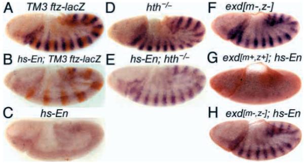
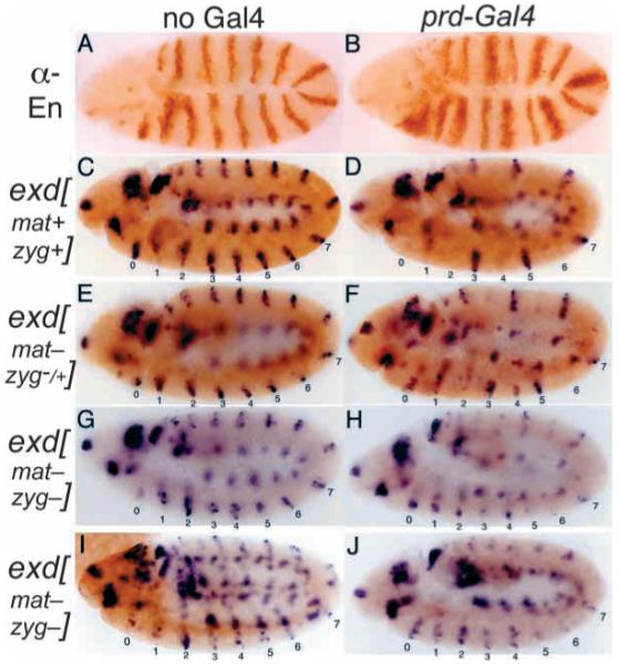
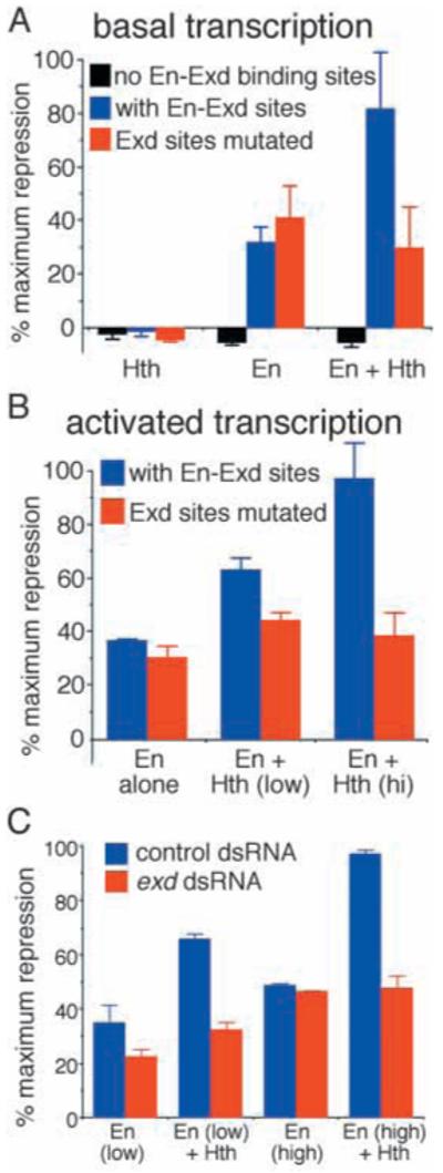
Engrailed is a key transcriptional regulator in the nervous system and in the maintenance of developmental boundaries in Drosophila, and its vertebrate homologs regulate brain and limb development. Here, we show that the functions of both of the Hox cofactors Extradenticle and Homothorax play essential roles in repression by Engrailed. Mutations that remove either of them abrogate the ability of Engrailed to repress its target genes in embryos, both cofactors interact directly with Engrailed, and both stimulate repression by Engrailed in cultured cells. We suggest a model in which Engrailed, Extradenticle and Homothorax function as a complex to repress Engrailed target genes. These studies expand the functional requirements for extradenticle and homothorax beyond the Hox proteins to a larger family of non-Hox homeodomain proteins.


Figures

Fig. 1
Fig. 1
Ubiquitously expressed Engrailed requires exd and hth to repress slp in vivo. Repression of slp by En is abrogated by loss of either hth or exd function. Embryos were collected for 1 hour, aged for 3.5 hours and heat shocked at 37°C for 10 minutes to induce En expression in hs-En embryos, which carry a hs-inducible transgene expressing full-length En). They were then aged for an additional 15 minutes, fixed and stained for slp RNA (dark blue, by in situ hybridization) and for β-galactosidase protein (A,B,D,E) (orange, anti-horseradish-peroxidase stain). lacZ is expressed from a ftz-lacZ transgene on the TM3 balancer chromosome. (A) Wild type (no hs-En, genotype TM3/hthP2 or TM3/TM3, which are indistinguishable). (B) hs-En, TM3 containing (TM3 carries a wild-type hth allele); notice the almost complete repression of slp by hs-En. (C) Control to show that TM3 itself does not affect repression of slp by hs-En (genotype hs-En; +/+, stained in parallel with the others). (D) An hth mutant (genotype hthP2/hthP2, distinguishable in the same population as in A by the absence of β-galactosidase staining). (E) An hth mutant with hs-En (genotype hs-En; hthP2/hthP2, inferred from the absence of β-galactosidase staining; this and the embryo in B were stained together in the same tube). Notice the weak repression of slp relative to B. (F-H) Embryos in F and H, designated ‘mat.exd-’ below (‘m-’ in the figure), were derived from germline clones in the mother that were homozygous for the null exd allele exd1; therefore, no maternally derived Exd was present. These females with exd- germlines were crossed either to wild-type males, to generate the embryo in F, or to males homozygous for a hs-En transgene, to generate the embryo in H. Wild-type females were crossed to the hs-En males to generate the embryo in G. (F) mat.exd-, without hs-En. (G) exd+, with hs-En. (H) mat.exd-, with hs-En. Notice the increase in slp RNA when Exd is missing (H versus G), indicating a requirement for Exd in order for En to efficiently repress slp. These embryos were also stained for the female-specific protein Sex-lethal, to indicate whether they were male or female. Because exd is on the X chromosome, females received a wild-type copy of the exd locus from their fathers, whereas males did not. The embryos shown were male, and so they received their only exd allele from their mother (‘z-’ or ‘z+’). A paternal-only contribution to exd function (that is, in females derived from females with exd- germlines) significantly increased the ability of hs-En to repress slp (data not shown).
Fig. 2
Fig. 2
Ectopically patterned En uses exd to repress wg in vivo. All embryos carry a UAS-en transgene. (A) Wild-type embryo stained for En. (B) Embryo carrying a prd-Gal4 driver transgene, in addition to UAS-en. Notice the anteriorly expanded alternate En stripes, which overlap alternate wg and slp stripes (Fig. 3). (C-J) Embryos were derived from either exd+/+ females (C,D) or from females with exd mutant germlines (E-J), and received either a wild-type exd allele (E,F) or no exd allele (G-J; exd is on the X chromosome) from their father. Thus exd[mat+ zyg+] refers to either a homozygous or hemizygous wild-type exd genotype; exd[mat- zyg-/+] refers to heterozygous females that lack a maternal contribution; and exd[mat- zyg-] refers to hemizygous mutants that also lack a maternal contribution. These embryos were double-stained for wg RNA by in situ hybridization and either for Exd (C-H), indicating whether they do or do not have a wild-type exd allele, or for a balancer marker (I,J; hb-lacZ, in brown, indicating absence of the prd-Gal4 driver transgene; lacZ-negative indicates the presence of the driver). Notice that, in exd wild-type embryos, wg is completely repressed by the ectopic En expression within alternate (even-numbered) stripes induced by prd-Gal4, whereas, in heterozygous exd embryos (which lack a maternal exd contribution), this repression is reduced. This effect was greater in embryos lacking all exd function (H,J). J is inferred to be exd mutant because of the weak and incomplete odd-numbered wg stripes that characterize them, as seen in H; H is inferred to contain prd-Gal4 because of the repression of even-numbered wg stripes in the abdomen, as seen in J. Notice the lack of repression of even-numbered stripes (particularly stripes 0, 2 and 4) in H and J, and, to a lesser extent, in F. Staining for En showed no difference in either the pattern or extent of ectopic expression between the wild-type and exd mutant populations (not shown).
Fig. 3
Fig. 3
Ectopically patterned Engrailed uses exd to repress slp in vivo. Embryos collected as in Fig. 2 were double stained for a balancer marker (hb-lacZ, in brown in C,D, indicating absence of the prd-Gal4 driver transgene; all others are lacZ negative, indicating that they carry the driver) and for slp RNA by in situ hybridization. Notice that, in exd wild-type embryos (A,B), slp is progressively repressed by the ectopic En expression within alternate stripes; in heterozygous exd embryos, which lack a maternal exd contribution (E,F), the repression is delayed and reduced; in embryos that lack all exd function both maternally and zygotically (G,H), this effect is stronger. Because repression is progressive, mutant embryos are shown that are at least as old as the corresponding wild-type controls.
Fig. 4
Fig. 4
Flies with exd mutations show dose-dependent interactions with ectopically patterned En in embryos. Embryos collected as in Figs 2 and 3 were allowed to develop to the end of embryogenesis, and cuticles were prepared and analyzed. Defects in abdominal denticle bands (mostly pairwise fusions of varying severity) were categorized and tabulated, and the results are shown in a stacked bar graph. As indicated by the percentage of defective cuticles in both the exd+/+ population and the population derived from exd mutant germline clones (data not shown), those embryos that did not receive a copy of the prd-Gal4 driver showed no defects other than those expected from the complete absence of exd function (embryonic cuticles from exd germline clones are completely rescued by one wild-type gene from the father). We did not attempt to analyze exd-null cuticles (which were clearly distinguishable from the exd heterozygotes) for effects of En ectopic expression induced by the driver. Rather, the denticle band fusions caused by ectopic En expression in the population that received a wild-type exd allele from their father were analyzed, and the graph shows the percentage of defects in each category among this population (which are exd+/-, as indicated in the key). Thus, the percentages add to 100% in each case because they include only those cuticles that showed defects caused by ectopic En expression that were of the indicated exd genotype (in each case, these represented the expected overall percentage of cuticles). Notice that when the maternal contribution of exd is removed and the zygotic contribution is simultaneously reduced by half (exd+/-), the overall severity of abdominal cuticle defects in the population caused by ectopic En expression is significantly reduced, indicating a substantial requirement for Exd in En function in the developing abdomen.
Fig. 5
Fig. 5
Engrailed interacts with Extradenticle and Homothorax in yeast, in vitro and in cultured cells. (A) En can interact with both Exd and Hth. In the yeast two-hybrid system, En was tested for interaction with full-length Exd, Hth and mouse Meis1. In each case, the protein listed first (or by itself) was expressed as a fusion with the Gal4 DNA-binding domain (DBD, in pAS2), whereas that listed second was fused with the Gal4 activation domain (in pACT2). En shows a strong signal with Exd and a weaker, but still specific, signal with both Hth and Meis1 (relative to these proteins either alone or in combination with the negative control P53, as indicated). Exd also generates a consistently strong signal with Meis1. (Hth produces a strong signal by itself when fused with the Gal4 DBD, so that similar experiments using it were uninformative.) (B) En interacts directly with both Exd and Meis1 in vitro and the three appear to form a co-complex. Glutathione-S-transferase (GST) fused to full-length En was produced and affinity purified from bacteria, then mixed with in vitro translated Exd (either labeled or unlabeled) and/or Meis1 (labeled), as indicated, and extracted from the mixture using glutathione agarose beads. Proteins captured by the beads were examined by SDS-PAGE and autoradiography. On the left, the single strong band migrates at the correct molecular weight to be full-length Exd. On the right, labeled Meis1 was mixed with either GST-En, GST-En plus unlabeled, in vitro translated Exd, GST alone or GST plus unlabeled Exd, as indicated. The prominent band migrates at the correct molecular weight to be authentic Meis1. (C) En interacts with Hth and Exd in cultured cells. Drosophila S2 cells were transfected with plasmids expressing Hth (panels I-IV, lanes 1-3), Hth plus En (panel V, lanes 1-3) or Hth plus En and His6Exd (‘tagExd’, lanes 4-6, all panels), and nuclear extracts were prepared (see Materials and Methods). Hth-specific antiserum (+, panels I and II) or preimmune serum control (-) was incubated with Protein-A/agarose beads and then with the nuclear extracts. His6-specific monoclonal antibodies (+, panels III-V) or nonspecific IgG control (-) were incubated with Protein-G/Sepharose beads and then with the nuclear extracts. ‘In’ indicates one-fifth of input extract (except panel III, where lanes 1 and 4 are shown at a shorter exposure to allow the En band to be clearly distinguished from a background band that is detected by the anti-En antiserum); P indicates a pellet (bead) fraction. Lanes 1-3 contain extract from the Hth-only (or Hth plus En, panel V only) transfection. Lanes 4-6 contain extract from Hth plus En and His6Exd transfection, analyzed by SDS-PAGE and western blotting, after incubation with control beads (-), anti-Hth (‘α-Hth’) beads (+, panels I and II) or anti-His6 (‘α-tag’) beads (+, panels III-V), followed by extensive washing, followed by detection of either En (panels I, III and V), Exd (panel II) or Hth (panel IV) with specific antisera. Notice that the background band (‘b.g.’) in panels III and V, which migrates faster than En and is present in both extracts, is not precipitated. This band does not appear in panel I because of the use of monoclonal anti-En antibody, whereas polyclonal anti-En antibody was used in panels III and V.
Fig. 6
Fig. 6
Hth and Exd enhance repression by En in cultured cells. Drosophila S2 cells were transfected with a target gene containing a CAT reporter, binding sites that are bound co-operatively by En and Exd in vitro (oligonucleotide sequence TCGAGTCAATTAAATGATCAATCAATTTCG); or, as indicated in the key, the same plasmid without these sites, or with a two-nucleotide change in an Exd core binding site (oligonucleotide sequence: TCGAGTCAATTAAAGCATCAATCAATTTCG) and activator binding sites. Cells were harvested after 66 hours and assayed for CAT and a co-transfected reference gene. The vertical axis shows results normalized to reference gene activity, as a percentage of the maximum repression observed in each experiment. Maximum repression was eightfold for basal transcription (A) and at least tenfold for activated transcription (B,C). Error bars show the range of values from two parallel transfections. (A) The reporter (either with or without En-Exd sites, or with mutated sites) and reference plasmids were co-transfected with either a Hth expression construct, an En expression construct, or both together. Bars below the baseline indicate the slight activation seen without binding sites. (B) The reporter (with or without Exd sites mutated as indicated in the key), activator and reference plasmids were co-transfected with an En expression construct either alone or in combination with a Hth expression construct at either a low or a higher concentration as indicated (see Materials and Methods for plasmid amounts and other transfection details). There was no effect of Hth alone on activated transcription. (C) The reporter, activator and reference plasmids were co-transfected with the En expression construct (at either a low or higher concentration) either alone or with the Hth expression construct, after a prior treatment of the cells with either control or Exd dsRNA, as indicated (see key).

Similar articles

Cited by

References

    1. Abu-Shaar M, Mann RS. Generation of multiple antagonistic domains along the proximodistal axis during Drosophila leg development. Development. 1998;125:3821–3830. - PubMed
    1. Abu-Shaar M, Ryoo HD, Mann RS. Control of the nuclear localization of Extradenticle by competing nuclear import and export signals. Genes Dev. 1999;13:935–945. - PMC - PubMed
    1. Abzhanov A, Holtzman S, Kaufman TC. The Drosophila proboscis is specified by two Hox genes, proboscipedia and Sex combs reduced, via repression of leg and antennal appendage genes. Development. 2001;128:2803–2814. - PubMed
    1. Akam M. The molecular basis for metameric pattern in the Drosophila embryo. Development. 1987;101:1–22. - PubMed
    1. Alexandre C, Vincent J-P. Requirements for transcriptional repression and activation by Engrailed in Drosophila embryos. Development. 2003;130:729–739. - PubMed

Publication types

MeSH terms

LinkOut - more resources