. 2003 Jan 7;100(1):330-5.
doi: 10.1073/pnas.222681699. Epub 2002 Dec 27.

Amyloid beta -protein (Abeta) assembly: Abeta 40 and Abeta 42 oligomerize through distinct pathways

Affiliations

Amyloid beta -protein (Abeta) assembly: Abeta 40 and Abeta 42 oligomerize through distinct pathways

Gal Bitan et al. Proc Natl Acad Sci U S A. .

Abstract

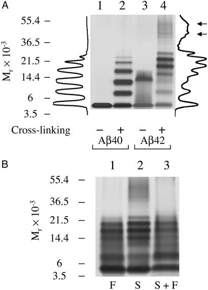
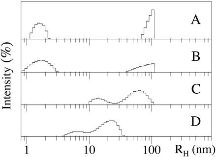
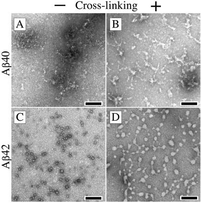
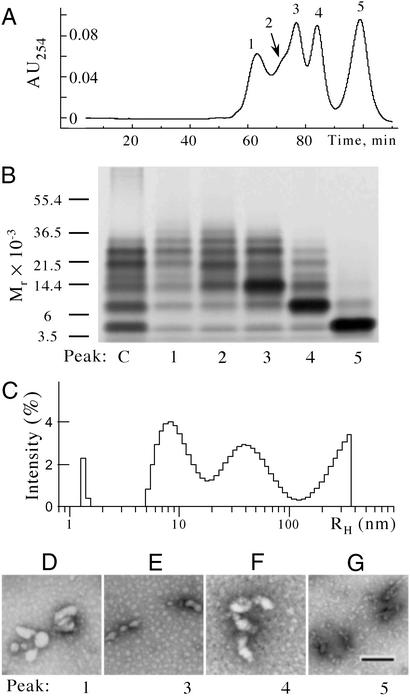
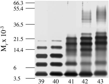
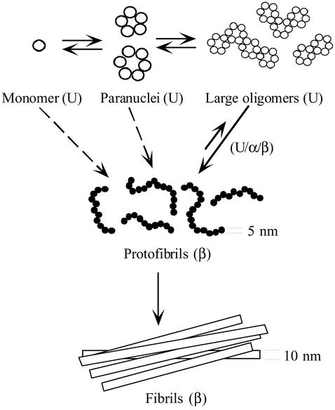
Amyloid beta-protein (Abeta) is linked to neuronal injury and death in Alzheimer's disease (AD). Of particular relevance for elucidating the role of Abeta in AD is new evidence that oligomeric forms of Abeta are potent neurotoxins that play a major role in neurodegeneration and the strong association of the 42-residue form of Abeta, Abeta42, with the disease. Detailed knowledge of the structure and assembly dynamics of Abeta thus is important for the development of properly targeted AD therapeutics. Recently, we have shown that Abeta oligomers can be cross-linked efficiently, and their relative abundances quantified, by using the technique of photo-induced cross-linking of unmodified proteins (PICUP). Here, PICUP, size-exclusion chromatography, dynamic light scattering, circular dichroism spectroscopy, and electron microscopy have been combined to elucidate fundamental features of the early assembly of Abeta40 and Abeta42. Carefully prepared aggregate-free Abeta40 existed as monomers, dimers, trimers, and tetramers, in rapid equilibrium. In contrast, Abeta42 preferentially formed pentamerhexamer units (paranuclei) that assembled further to form beaded superstructures similar to early protofibrils. Addition of Ile-41 to Abeta40 was sufficient to induce formation of paranuclei, but the presence of Ala-42 was required for their further association. These data demonstrate that Abeta42 assembly involves formation of several distinct transient structures that gradually rearrange into protofibrils. The strong etiologic association of Abeta42 with AD may thus be a result of assemblies formed at the earliest stages of peptide oligomerization.


Figures

Figure 1
Figure 1
PICUP reveals distinct oligomer distributions for Aβ40 and Aβ42. LMW Aβ40 and Aβ42 were cross-linked immediately after preparation by SEC and analyzed by SDS/PAGE/silver staining. (A) Noncross-linked (lanes 1 and 3) or cross-linked (lanes 2 and 4) Aβ40 and Aβ42. Intensity profiles of lanes 2 (cross-linked Aβ40) and 4 (cross-linked Aβ42) are shown (left and right of the gel, respectively). These profiles were generated by using ONE-DSCAN (Scanalytics, Fairfax, VA). Arrows next to the intensity profile of cross-linked Aβ42 indicate intensity maxima corresponding to presumptive dodecamer and octadecamer species. (B) PICUP chemistry was performed on LMW Aβ42 samples immediately after preparation by filtration (“F”) through a 10,000 Mr cut-off filter (lane 1), SEC (“S,” lane 2), or SEC followed by filtration (“S + F,” lane 3). Positions of molecular weight standards (A and B) are shown on the left. The gels are representative of those obtained in each of at least three independent experiments.
Figure 2
Figure 2
Analysis of the oligomer size distributions of Aβ by DLS. Representative DLS spectra are shown of LMW Aβ40 isolated by SEC (A) or filtration (B). Large particles (>100 nm) were not included in the measurement window, resulting in the truncation of the peaks in that region. Representative DLS spectra are shown of LMW Aβ42 isolated by SEC (C) or filtration (D). The data are representative of those obtained in each of at least three independent experiments.
Figure 3
Figure 3
Morphologic analysis of cross-linked LMW Aβ. LMW Aβ40 (A and B) or Aβ42 (C and D) were isolated by using SEC. Aliquots of noncross-linked (A and C) or cross-linked (B and D) peptide were spotted on glow-discharged, carbon-coated grids, stained with uranyl acetate, and examined by EM. (Bars = 100 nm.) The images are representative of those in each of at least three independent experiments.
Figure 4
Figure 4
Comparative analysis of Aβ42 oligomer morphology and size distribution. (A) Cross-linked Aβ42 was fractionated by SEC and five peaks were collected (indicated as 1–5). SDS/PAGE (B), DLS (C), and EM (D) then were done on the five peaks obtained to characterize and correlate morphologic and oligomer size distribution data. (B) SDS/PAGE/silver stain analysis. Positions of molecular weight standards are shown on the left. (C) DLS analysis of peak 1. (D–G) EM images of peaks 1, 3, 4, and 5, respectively. (Bar = 100 nm.) The data shown are representative of those obtained in each of at least three independent experiments.
Figure 5
Figure 5
C-terminal length-dependence of Aβ oligomerization. SEC-isolated LMW Aβ39, Aβ40, Aβ41, Aβ42, and Aβ43 were cross-linked individually and analyzed by SDS/PAGE. Positions of molecular weight standards are shown on the left. The gel is representative of three independent experiments.
Figure 6
Figure 6
A simple model of Aβ42 assembly. Monomers rapidly oligomerize into paranuclei. Paranuclei themselves then can oligomerize to form larger, beaded structures. Paranuclei are the initial, and minimal, structural unit from which Aβ42 assemblies evolve. The equilibrium between monomer and paranucleus is rapid, as evidenced by the fact that paranuclei are detectable immediately after peptide dissolution. The conversion to protofibrils is slower (12), but is also reversible. Monomers, paranuclei, and large oligomers are predominately unstructured (U), but do contain β-sheet/β-turn (β) and helical (α) elements (see text). Protofibril formation involves substantial conformational rearrangements, during which unstructured, α-helix, and β-strand elements (U/α/β) transform into predominately β-sheet/β-turn structures. Protofibrils may form through the oligomerization of monomers into paranuclei, paranucleus self-association to form larger oligomers, and then maturation of these large oligomers into protofibrils. This “linear” pathway may not be the only one. Direct addition of monomers or paranuclei (dotted arrows) to protofibrils or fibrils cannot be ruled out. The final step in the overall pathway is protofibril maturation into fibrils, a process that appears irreversible, at least kinetically (38). The diameters of the protofibrils and fibrils are indicated, but the structures in the scheme are not drawn to scale.

References

    1. Hardy J A, Higgins G A. Science. 1992;256:184–185. - PubMed
    1. Selkoe D J. J Neuropathol Exp Neurol. 1994;53:438–447. - PubMed
    1. Hardy J. Proc Natl Acad Sci USA. 1997;94:2095–2097. - PMC - PubMed
    1. Glenner G G, Wong C W. Biochem Biophys Res Commun. 1984;120:885–890. - PubMed
    1. Masters C L, Simms G, Weinman N A, Multhaup G, McDonald B L, Beyreuther K. Proc Natl Acad Sci USA. 1985;82:4245–4249. - PMC - PubMed

Publication types

MeSH terms