Low frequency of FAS mutations in Reed-Sternberg cells of Hodgkin's lymphoma
- PMID: 12507887
- PMCID: PMC1851130
- DOI: 10.1016/S0002-9440(10)63795-9
Low frequency of FAS mutations in Reed-Sternberg cells of Hodgkin's lymphoma
Abstract
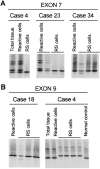
Reed-Sternberg (RS) cells, the neoplastic elements of Hodgkin's lymphoma (HL), usually lack B-cell receptor expression. Normal germinal center B cells, with lack of or low-affinity B-cell receptor expression, are eliminated via FAS-induced apoptosis. RS cells express FAS, but are rescued from apoptosis by a transforming event. It is known that HL-derived cell lines are resistant to FAS-mediated apoptosis. To investigate potential causes for this resistance, FAS mutations and c-FLIP expression were studied in four HL-derived cell lines and 20 cases of HL. L1236 was found to have a splice donor site mutation in intron 7 that resulted in an aberrantly spliced FAS transcript. Screening of microdissected RS cells revealed loss of heterozygosity for a known exon 7 polymorphism in two of six informative cases indicating loss of one FAS allele. In one of the two cases with loss of heterozygosity a hemizygous mutation was detected in exon 9. c-FLIP expression was observed in all HL cell lines and in RS cells of all HL cases. Our data show that FAS mutations are rare and suggest that overexpression of c-FLIP, which was present in all cases, is involved in the resistance to FAS-mediated apoptosis.
Figures


Similar articles
-
Constitutive expression of c-FLIP in Hodgkin and Reed-Sternberg cells.Am J Pathol. 2002 Apr;160(4):1521-8. doi: 10.1016/S0002-9440(10)62578-3. Am J Pathol. 2002. PMID: 11943736 Free PMC article.
-
Expression of the cellular FLICE-inhibitory protein (c-FLIP) protects Hodgkin's lymphoma cells from autonomous Fas-mediated death.Proc Natl Acad Sci U S A. 2004 Apr 27;101(17):6611-6. doi: 10.1073/pnas.0400765101. Epub 2004 Apr 19. Proc Natl Acad Sci U S A. 2004. PMID: 15096587 Free PMC article.
-
c-FLIP mediates resistance of Hodgkin/Reed-Sternberg cells to death receptor-induced apoptosis.J Exp Med. 2004 Apr 19;199(8):1041-52. doi: 10.1084/jem.20031080. Epub 2004 Apr 12. J Exp Med. 2004. PMID: 15078899 Free PMC article.
-
Hodgkin's lymphoma: the role of cell surface receptors in regulation of tumor cell fate.Exp Oncol. 2010 Dec;32(4):214-23. Exp Oncol. 2010. PMID: 21270747 Review.
-
Molecular biology of Hodgkin's and Reed/Sternberg cells in Hodgkin's lymphoma.Int J Cancer. 2006 Apr 15;118(8):1853-61. doi: 10.1002/ijc.21716. Int J Cancer. 2006. PMID: 16385563 Review.
Cited by
-
Analysis of the mutational landscape of classic Hodgkin lymphoma identifies disease heterogeneity and potential therapeutic targets.Oncotarget. 2017 Nov 30;8(67):111386-111395. doi: 10.18632/oncotarget.22799. eCollection 2017 Dec 19. Oncotarget. 2017. PMID: 29340061 Free PMC article.
-
cIAP2 is highly expressed in Hodgkin-Reed-Sternberg cells and inhibits apoptosis by interfering with constitutively active caspase-3.J Mol Med (Berl). 2006 Feb;84(2):132-41. doi: 10.1007/s00109-005-0003-7. Epub 2005 Nov 25. J Mol Med (Berl). 2006. PMID: 16308685
-
Genetic lesions and targeted therapy in Hodgkin lymphoma.Ther Adv Hematol. 2023 Jan 12;14:20406207221149245. doi: 10.1177/20406207221149245. eCollection 2023. Ther Adv Hematol. 2023. PMID: 36654739 Free PMC article. Review.
-
Hodgkin Lymphoma: A Special Microenvironment.J Clin Med. 2021 Oct 12;10(20):4665. doi: 10.3390/jcm10204665. J Clin Med. 2021. PMID: 34682791 Free PMC article. Review.
-
Immune Checkpoint Inhibition in Classical Hodgkin Lymphoma: From Early Achievements towards New Perspectives.J Oncol. 2019 May 7;2019:9513701. doi: 10.1155/2019/9513701. eCollection 2019. J Oncol. 2019. PMID: 31205470 Free PMC article. Review.
References
-
- Küppers R, Rajewsky K: The origin of Hodgkin and Reed Sternberg cells in Hodgkin’s disease. Annu Rev Immunol 1998, 16:471-493 - PubMed
-
- Rajewsky K: Clonal selection and learning in the antibody system. Nature 1996, 381:751-758 - PubMed
-
- Chapman ALN, Rickinson AB: Epstein-Barr virus in Hodgkin’s disease. Ann Oncol 1995, 9:5-16 - PubMed
-
- Jarrett RF, MacKenzie J: Epstein-Barr virus and other candidate viruses in the pathogenesis of Hodgkin’s disease. Semin Hematol 1999, 36:260-269 - PubMed
Publication types
MeSH terms
Substances
LinkOut - more resources
Full Text Sources
Medical
Research Materials
Miscellaneous

