. 2003 Jan;162(1):37-45.
doi: 10.1016/S0002-9440(10)63796-0.

Quantitative gene expression analysis reveals transition of fetal liver progenitor cells to mature hepatocytes after transplantation in uPA/RAG-2 mice

Affiliations

Quantitative gene expression analysis reveals transition of fetal liver progenitor cells to mature hepatocytes after transplantation in uPA/RAG-2 mice

Tobias Cantz et al. Am J Pathol. 2003 Jan.

Abstract

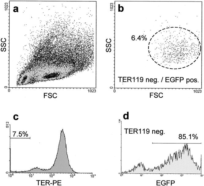
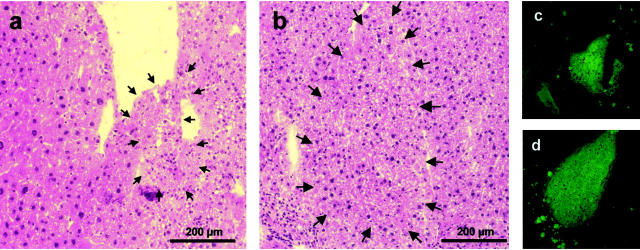
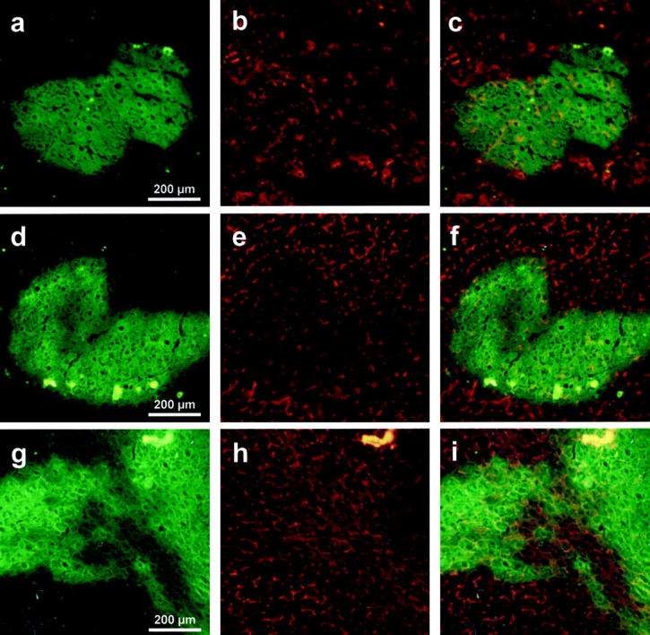
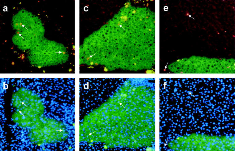
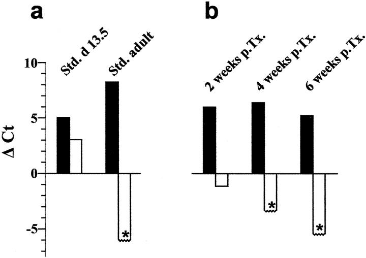
Therapies for liver diseases with stem and progenitor cells will require a detailed knowledge of the molecular mechanisms driving the in vivo differentiation process toward adult hepatic tissue. We applied quantitative gene expression methods to analyze the differentiation process of fetal liver progenitor cells after transplantation into an animal model of liver regeneration. Enhanced green fluorescent protein (EGFP)-transgenic liver progenitor cells were isolated from fetal mouse liver at stage embryonic day 13.5 and transplanted into uPA/RAG-2 mice. Two, 4, and 6 weeks after cell transplantation cryosections of liver tissue were analyzed for EGFP-positive regeneration nodules. RNA from laser-microdissected EGFP-positive tissue was isolated and used as template for quantitative real-time reverse transcriptase-polymerase chain reaction. Phenotypic differentiation was analyzed by staining of the canalicular marker enzyme dipeptidyl-peptidase IV. Proliferation in regenerative nodules and surrounding tissue was monitored with the BrdU incorporation assay. Alpha fetoprotein gene expression had already decreased 2 weeks after transplantation in EGFP-positive regeneration nodules compared to pretransplantation values and was not detectable after 4 and 6 weeks, whereas albumin slightly increased in transplanted cells indicating differentiation into a mature phenotype. The dipeptidyl-peptidase IV antigen was associated with some liver progenitor cells 2 weeks after transplantation and in virtually all cells after 4 and 6 weeks. Cell proliferation index in transplanted cells was maximally increased (4.8% BrdU-positive cells) after 2 weeks and decreased (0.4%) after 6 weeks to normal levels. Our results demonstrate that gene expression in liver progenitor cells changes from fetal to adult phenotype within 4 to 6 weeks after transplantation despite ongoing proliferation of the transplanted cells in a mouse model of liver regeneration. Quantitative gene expression profiles as shown here will have important implications in our understanding of the in vivo differentiation process of stem cells.


Figures

Figure 1.
Figure 1.
FACS analysis of day 13.5 fetal liver progenitor cells of EGFP-C57BL/6 mice. For fluorescent cell cytometry analysis of cell suspension derived from fetal liver (embryonic day 13.5) staining with the erythrocyte marker TER119 was performed and vitality was determined by propidium iodide (PI) exclusion. Cells (n = 100,000) were collected and PI-negative cells (97.5%, data not shown) were analyzed by the foreword/sideward scatter (a). Most of the cells in fetal livers were of hematopoietic origin with a high percentage of erythrocytes (92.5% TER-positive) that did not express EGFP (c). Because heterozygous mice were used for the mating, there are some embryos with EGFP-negative liver progenitor cells. From the remaining 7.5% TER-negative cells the majority was EGFP-positive (85.1%), demonstrating a small number of cells derived from EGFP-negative fetal livers only (d). The overall percentage of TER119-negative EGFP-positive cells was 6.4%. These cells contained the fraction of liver progenitor cells and were distributed typically in the FSC/SSC blot (b).
Figure 2.
Figure 2.
Regeneration nodules after transplantation of EGFP-positive fetal liver progenitor cells in uPA/RAG-2 mice. Five-μm cryosections of uPA/RAG-2 mouse livers were stained with hemalaun-eosin and consecutive slides were analyzed for EGFP fluorescence after EGFP-positive fetal liver progenitor cell transplantation. Two weeks (a and c) after transplantation, small EGFP-positive regeneration nodules (arrows) are detectable between normal endogenously regenerated (a, left) and uPA-damaged liver tissue (a, right). After 4 weeks (b and d) the damaged liver tissue nearly was diminished and the nodules have become larger, whereby the liver morphology in the H&E staining developed a normal pattern (b). On the right part of this nodule there is a demarcation line, which is probably a result of the freezing, fixation, and staining procedure.
Figure 3.
Figure 3.
Immunofluorescence analysis of the DPPIV antigen in EGFP-positive regeneration nodules. Five-μm cryosections from uPA/RAG2-mouse livers were analyzed 2 weeks (no. 1246, a–c), 4 weeks (no. 1255, d–f), and 6 weeks (no. 1308, g–i) after transplantation of EGFP fetal liver cells. For analysis of the fluorescent (green) regeneration nodules co-staining of the hepatic marker enzyme DPPIV was performed (red fluorescence), illustrating the canalicular membranes of polarized hepatocytes (b, e, h). The overlay of the green fluorescent regeneration nodules with the red DPPIV signal shows the presence of few polarized hepatocytes 2 weeks after transplantation (c), much more after 4 weeks (f), and a normal pattern after 6 weeks (i), demonstrating the developing liver architecture and the integration of the regenerative tissue.
Figure 4.
Figure 4.
Proliferation activity index in EGFP-positive regeneration nodules. Specimens of fetal liver progenitor cell-derived regeneration nodules (green fluorescence) in uPA/RAG2-mouse liver were analyzed 2 weeks (no. 1246, a and b) and 4 weeks (no. 1255, c–f) after transplantation for BrdU incorporation (red nuclear fluorescence, arrows in a, c, e). The ratio of the number of BrdU versus the total number of 4,6-diamidino-2-phenylindole staining of all nuclei (b, d, f) was calculated, as demonstrated by the corresponding arrows. Some unspecific autofluorescence in the diseased liver tissue appears in the green and red channel resulting in yellow spots around the regeneration nodules. Increased proliferation activity is shown in regeneration nodules at 2 weeks (a and b) and at a lower degree also at 4 weeks (c and d) after fetal liver progenitor cell transplantation, whereas in the surrounding endogenously regenerated liver tissue (e and f) only few proliferating cells are detectable (see also Table 1 ▶ ).
Figure 5.
Figure 5.
Expression profiles of albumin and AFP in liver progenitor cell differentiation. The albumin (black bars) and AFP expression (white bars) was normalized to GAPDH by subtracting their Ct values from the GAPDH Ct value (see Table 2 ▶ ). For calculating GAPDH-AFP Ct differences the detection limit of AFP expression was set at a value of 38 (asterisks). For each experimental group the means were calculated and shown in this figure. In the control experiments (a) the standard cDNA of day 13.5 fetal liver and adult liver showed an albumin expression of +5.05 and +8.25, respectively. In contrast the AFP expression was +3.05 in the fetal liver cDNA and under the detection limit (<−5.95) in adult liver cDNA. Two, 4, and 6 weeks after transplantation (b) the expression profiles of the transplanted liver progenitor cells altered toward mature hepatocytes. The albumin expression remained constant (+6.00, +6.40, +5.23), whereas the AFP-expression decreased gradually (−1.13 after 2 weeks) under the detection limit (<−3.30 and <−5.4 after 4 and 6 weeks, respectively).

References

    1. Cascio SM: Novel strategies for immortalization of human hepatocytes. Artif Organs 2001, 25:529-538 - PubMed
    1. Fox IJ, Chowdhury JR, Kaufman SS, Goertzen TC, Chowdhury NR, Warkentin PI, Dorko K, Sauter BV, Strom SC: Treatment of the Crigler-Najjar syndrome type I with hepatocyte transplantation. N Engl J Med 1998, 338:1422-1426 - PubMed
    1. Bilir BM, Guinette D, Karrer F, Kumpe DA, Krysl J, Stephens J, McGavran L, Ostrowska A, Durham J: Hepatocyte transplantation in acute liver failure. Liver Transpl 2000, 6:32-40 - PubMed
    1. De Vree JM, Ottenhoff R, Bosma PJ, Smith AJ, Aten J, Oude Elferink RP: Correction of liver disease by hepatocyte transplantation in a mouse model of progressive familial intrahepatic cholestasis. Gastroenterology 2000, 119:1720-1730 - PubMed
    1. Overturf K, Al Dhalimy M, Ou CN, Finegold M, Grompe M: Serial transplantation reveals the stem-cell-like regenerative potential of adult mouse hepatocytes. Am J Pathol 1997, 151:1273-1280 - PMC - PubMed

Publication types

MeSH terms

Substances