. 2003 Jan;162(1):139-50.
doi: 10.1016/S0002-9440(10)63805-9.

Experimental autoimmune encephalomyelitis (EAE) in CCR2(-/-) mice: susceptibility in multiple strains

Affiliations

Experimental autoimmune encephalomyelitis (EAE) in CCR2(-/-) mice: susceptibility in multiple strains

Stefanie Gaupp et al. Am J Pathol. 2003 Jan.

Abstract

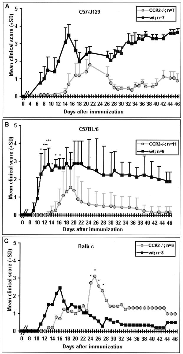
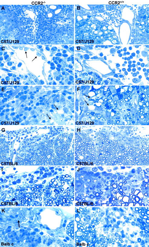
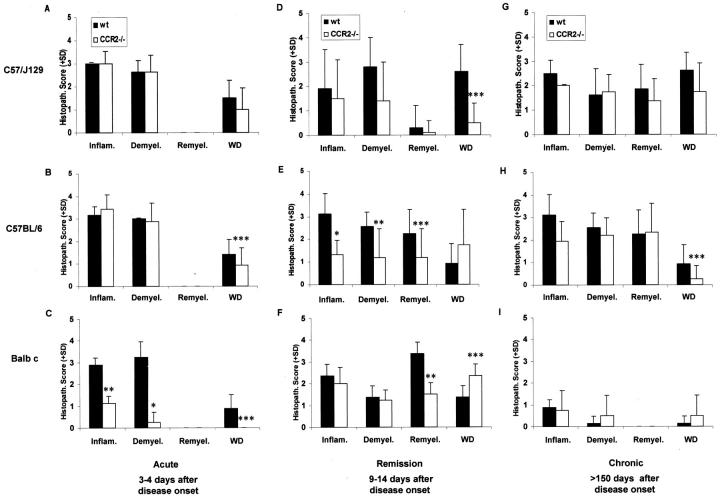
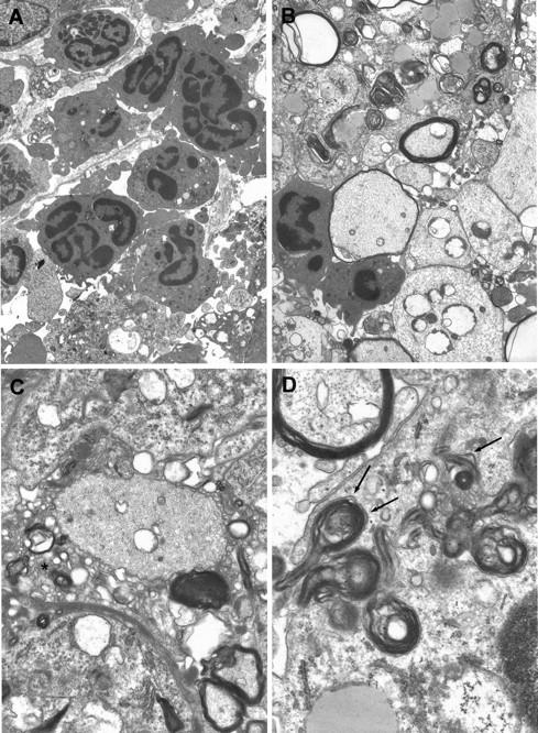
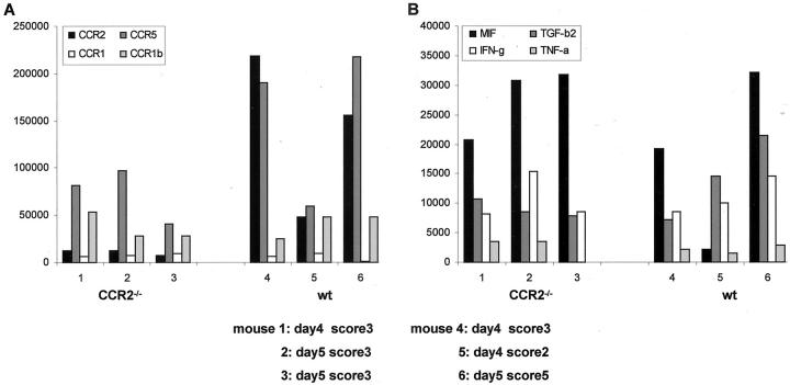
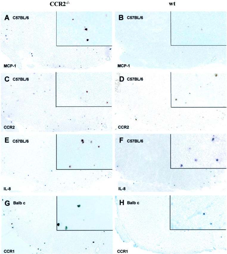
Chemokines are low molecular weight cytokines which act as chemoattractants for infiltrating cells bearing appropriate receptors (CCR) to sites of inflammation. It has been proposed that CCR2 on monocytes is responsible for their recruitment into the central nervous system (CNS) in experimental autoimmune encephalomyelitis (EAE), a model for multiple sclerosis, and two previous reports have described resistance of CCR2(-/-) mice to EAE. The present study examined three different mouse strains with CCR2 deletions for susceptibility to EAE. Animals were studied up to 4 months post-sensitization and were examined by neuropathology, RNase protection assay, in situ hybridization, and in vitro assays. All three strains were found to be susceptible to EAE: C57BL/6 x J129 and Balb c strains, 100%; and C57BL/6, 67%. Unusual in CNS lesions of CCR2(-/-) mice was an overabundance of neutrophils versus monocytes in wild-type animals. An attempt of the immune system to develop compensatory mechanisms for the lack of CCR2 was evidenced by a corresponding increase in mRNA for other chemokines and CCR. Inasmuch as neutrophils replaced monocytes and led to demyelination, our findings support the concept that promiscuity of chemokines and CCR was able to surmount the deletion of CCR2, still resulting in full expression of this autoimmune disease.


Figures

Figure 1.
Figure 1.
CCR2−/− mice from all three strains were susceptible to EAE. A: C57/J129; note the relapsing course of MOG35–55-induced EAE. B: C57/BL6; note the monophasic course in mice immunized with MOG35–55. C: Balb c; note monophasic course after immunization with guinea pig crude myelin. Both C57 strains showed delayed onset and reduced severity while Balb c displayed delayed onset with clinical scores comparable to wt mice. Clinical scores were assessed daily after immunization on day 0. Mean values and SD are indicated; P values refer to comparison between ko and wt mice. *, P < 0.001; **, P < 0.01; ***, P < 0.05.
Figure 2.
Figure 2.
Typical acute EAE lesions in ko and wt. A–F: C57/J129 mice. Note that the inflammatory infiltrates are composed predominantly of neutrophils in CCR2−/− mice (A and C) versus monocytes in wt animals (B and D). Axonal damage is apparent as ballooned and condensed nerve fibers (WD), seen mainly in wt animals (B, D, F), and rarely in CCR2−/− (A, C, E). Arrows in C indicate high endothelial venules in ko animals. Demyelinated fibers were found in both CCR2−/− and wt animals (arrows in E and F). A: Magnification, ×250, B: Magnification, ×250, C: Magnification, ×625, D: Magnification, ×625, E: Magnification, ×625, F: Magnification, ×625. G–J: C57BL/6 mice displayed similar inflammatory infiltrates comprising neutrophils, more so in CCR2−/− (G, I), monocytes, macrophages, and lymphocytes. More severe demyelination was observed in the spinal cord of wt mice (H, J). G: Magnification, ×250, H: Magnification, ×250, I: Magnification, ×625, J: Magnification, ×625. K and L: Balb c CCR2−/− mice (K) mice displayed a preponderance of neutrophils, while wt lesions contained more monocytes (L). Note the affected root entry zone at arrow in (K). K: Magnification, ×625, L: Magnification, ×625.
Figure 3.
Figure 3.
Histopathological differences between CCR2−/− and wt mice with disease progression. A–C: Acute EAE. Balb c CCR2−/− mice revealed decreased pathology compared to wt. D–F: Remission. CCR2−/− mice of both C57 strains displayed less pathology (D and E), which was less prominent in Balb c ko animals (F). Interestingly, CCR2−/− mice with a C57/J129 background showed decreased axonal damage (D), in contrast to C57BL/6 (E) and Balb c CCR2 ko mice (F). G–I. Chronic EAE. Balb c mice showed less pathology, but increased demyelination and WD in CCR2−/− mice (I), in contrast to both CCR2−/− from C57 strains (G and H). Data represent average histological scores of three animals (C57 strains) or a single animal (Balb c), evaluated from semi-thin cross sections of lower spinal cord (four levels, eight slides). *, ± P < 0.001; **, P < 0.01; ***, P < 0.05
Figure 4.
Figure 4.
Ultrastructure of CCR2−/− mice showed features typical of wt acute EAE. Section of lumbar spinal cord from a CCR2−/− C57/J129 mouse (clinical score 3), day 4 after disease onset, is depicted. A: Neutrophils are seen within the leptomeningial compartment and the CNS parenchyma. One neutrophil can be seen traversing the glia limitans (arrows). Magnification, ×4400. B: Demyelinated axons (lower right) are shown and one nerve fiber displays an attenuated sheath (left center) flanked by two neutrophils. Elsewhere, macrophages contain myelin debris. Magnification, ×5300. C: The process of demyelination in CCR2−/− mice was intact. Disrupted myelin forms a myelin network around the demyelinated axon (center). Magnification, ×1300. D: Uptake of myelin by a macrophage occurs via its attachment to clathrin-coated pits (arrows) on the cell surface, a phenomenon known as receptor-mediated phagocytosis. Magnification, ×2500.
Figure 5.
Figure 5.
RPA quantification of chemokine/chemokine receptor/cytokine mRNA expression in CNS tissue of CCR2−/− and wt C57/J129 mice with acute EAE. Intensity of the signal for protected bands was determined by phosphoimaging. Data are expressed as a ratio of the band of interest to the sum of signals for L32 and GAPDH. In ko animals, mRNA transcripts of CCR2 and CCR5 in CNS tissue were decreased (A). However, mRNA expression of MIF and cytokines, like IL-1, TGF-β, IFN-γ, and TNF-α, was comparable in CCR2−/− and wt mice (B).
Figure 6.
Figure 6.
ISH for mRNA expression for chemokine receptors and their ligands in CCR2−/− and wt revealed increased MCP-1 mRNA expression in lumbar spinal cord of CCR2−/− (A) versus wt (B) C57BL/6 mice, while numbers of cells expressing CCR2 mRNA were comparable in both groups (C and D). The number of IL-8-expressing cells was higher in CCR2−/− (E) compared to wt (F) mice. Balb c CCR2 −/− mice displayed a higher mRNA expression for CCR1 (G), compared to corresponding wt mice (H). Magnification, ×100; insets: magnification, ×450.

References

    1. Raine CS: Biology of disease. The analysis of autoimmune demyelination: its impact upon multiple sclerosis. Lab Invest 1984, 50:608-635 - PubMed
    1. Steinman L: Assessment to the utility of animal models for MS and demyelinating disease in the design of rational therapy. Neuron 1999, 24:511-514 - PubMed
    1. Raine CS: The lesion in multiple sclerosis and chronic relapsing experimental allergic encephalomyelitis: a structural comparison. Raine CS McFarland HF Tourtellotte WW eds. Multiple Sclerosis: Clinical and Pathogenetic Basis. 1997:pp 243-286 Chapman and Hall, London
    1. Cross AH, Cannella B, Brosnan CF, Raine CS: Homing to central nervous system vasculature by antigen-specific lymphocytes: I. localization of 14C-labeled cells during acute, chronic, and relapsing experimental allergic encephalomyelitis. Lab Invest 1990, 63:162-170 - PubMed
    1. Baggiolini M, Dahinden CA: CC chemokines in allergic inflammation. Immunol Today 1994, 15:127-133 - PubMed

Publication types

MeSH terms

LinkOut - more resources