. 2003 Jan;162(1):203-11.
doi: 10.1016/S0002-9440(10)63811-4.

Arginase 1 overexpression in psoriasis: limitation of inducible nitric oxide synthase activity as a molecular mechanism for keratinocyte hyperproliferation

Affiliations

Arginase 1 overexpression in psoriasis: limitation of inducible nitric oxide synthase activity as a molecular mechanism for keratinocyte hyperproliferation

Daniela Bruch-Gerharz et al. Am J Pathol. 2003 Jan.

Abstract

Abnormal proliferation of keratinocytes in the skin appears crucial to the pathogenesis of psoriasis, but the underlying mechanisms remain unknown. Nitric oxide (NO), released from keratinocytes at high concentrations, is considered a key inhibitor of cellular proliferation and inducer of differentiation in vitro. Although high-output NO synthesis is suggested by the expression of inducible NO synthase (iNOS) mRNA and protein in psoriasis lesions, the pronounced hyperproliferation of psoriatic keratinocytes may indicate that iNOS activity is too low to effectively deliver anti-proliferative NO concentrations. Here we show that arginase 1 (ARG1), which substantially participates in the regulation of iNOS activity by competing for the common substrate L-arginine, is highly overexpressed in the hyperproliferative psoriatic epidermis and is co-expressed with iNOS. Expression of L-arginine transporter molecules is found to be normal. Treatment of primary cultured keratinocytes with Th1-cytokines, as present in a psoriatic environment, leads to de novo expression of iNOS but concomitantly a significant down-regulation of ARG1. Persistent ARG1 overexpression in psoriasis lesions, therefore, may represent a disease-associated deviation from normal expression patterns. Furthermore, the culturing of activated keratinocytes in the presence of an ARG inhibitor results in a twofold increase in nitrite accumulation providing evidence for an L-arginine substrate competition in human keratinocytes. High-output NO synthesis is indeed associated with a significant decrease in cellular proliferation as shown by down-regulation of Ki67 expression in cultured keratinocytes but also in short-term organ cultures of normal human skin. In summary, our data demonstrate for the first time a link between a human inflammatory skin disease, limited iNOS activity, and ARG1 overexpression. This link may have substantial implications for the pathophysiology of psoriasis and the development of new treatment strategies.


Figures

Figure 1.
Figure 1.
RT-PCR analysis of keratoma biopsies from patients with psoriasis (n = 10), basal cell carcinoma (n = 3), and from healthy volunteers (n = 2). RNA extracted from skin samples was reverse-transcribed and analyzed by PCR with primers specific for ARG1 (550 bp, lane 1), iNOS (394 bp, lane 2), CAT-2 (457 bp, lane 3), and G3PDH sequences (416 bp, lane 4). Case numbers are shown above the lanes. ARG1 and iNOS mRNA are present in all biopsies from psoriasis patients (lines 1 to 5; shown are 5 of 10 psoriasis specimens). No iNOS-specific signal was found in skin specimens from healthy volunteers (lines 6 to 8; shown are three of five normal skin specimens), and a weak signal for ARG1 is seen after six additional amplification cycles only (not shown). In basal cell carcinomas a similar high level of ARG1 mRNA expression was observed (lines 9 and 10; shown are two of five basal cell carcinomas) but no iNOS expression. Conversely, CAT-2 gene expression was found in all biopsies investigated.
Figure 2.
Figure 2.
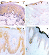
Co-expression of iNOS and ARG1 in skin specimens of psoriasis by immunohistochemistry. In immunolabeling of cryostat sections brown signals are obtained with blue nuclei because of hematoxylin counterstaining. Both ARG1 and iNOS protein are present in the hyperproliferative psoriatic epidermis. In A the psoriatic skin ARG1 immunoreactivity is found all over the epidermis, whereas in B iNOS is localized to the basal and suprabasal cell layers of psoriatic epidermis. Note the prominent staining of both enzymes in the basal layers of the hyperproliferative epidermis indicating that ARG1 and iNOS are expressed simultaneously. C: Control studies performed with isotype-matched control antibodies were uniformly negative. D: In normal skin specimens a weak ARG1-specific signal is found all over the epidermis.
Figure 3.
Figure 3.
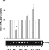
RT-PCR detection of iNOS, ARG1, CAT-1 and CAT-2 mRNA in resident or cytokine-challenged cultured human keratinocytes. Primary keratinocytes were incubated for 24 hours in the presence or absence of 1000 U/ml of γ-interferon, 1000 U/ml of tumor necrosis factor-α, and 1000 U/ml of interleukin-1β. Resident keratinocytes (r) constitutively express ARG1 mRNA and ARG2 mRNA as well as the cationic amino acid transporters CAT-1 and CAT-2. Cytokine activation of primary keratinocytes (a) leads to de novo expression of iNOS mRNA, up-regulation of CAT-2 mRNA by a factor of 1.56 ± 0.1 (*, P < 0.02) and down-regulation of ARG1 mRNA to a level of 45 ± 12% (*, P < 0.001) of resident cells. CAT-1 and ARG2 were not significantly influenced at their mRNA expression level. The figure shows data compiled from three experiments. Relative signal intensities (ie, amplification products per housekeeping gene) were set to 100%, with the exception of iNOS.
Figure 4.
Figure 4.
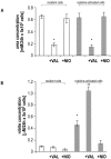
Inhibition of ARG activity during cytokine challenge significantly increases NO production. A: ARG activity as determined by urea concentrations in culture supernatants is high in resident cells. Within 24 hours of cytokine activation ARG activity remains unchanged. Supplementation with l-valine (VAL, 10 mmol/L) inhibits ARG activity in resident (white bars) as well as in activated cells (gray bars) (*, P < 0.005), whereas a NOS inhibitor (NIO, 0.25 mmol/L) does not influence ARG activity. B: Resident cells produce background levels of nitrite only (white bars), and after 24 hours of cytokine-challenge (gray bars) the increase in nitrite concentrations (*, P < 0.001 as compared to resident cells) demonstrates iNOS activity. Inhibition of NOS decreases nitrite production to background levels, but supplementation with l-valine increases nitrite formation by a factor of 2.1 ± 0.1 (**, P > 0.01) as compared to activated cells. Bars represent the mean ± SD of three experiments.
Figure 5.
Figure 5.
Endogenous and exogenous NO down-regulates proliferation in organ cultures of human skin and cultured keratinocytes. A: Maintenance of normal skin specimens for 24 hours in the presence of DETA/NO (1 mmol/L) leads to a strong reduction of Ki67 mRNA expression (black bar) to 8 ± 2.5% of the control (*, P < 0.001). The figure shows data compiled from three experiments. Relative amplification products are normalized to Ki67 expression of control cultures (r). B: Primary keratinocyte cultures (r) were activated with proinflammatory cytokines as in Figure 3 ▶ for 24 hours in the presence (a + NIO) or absence (a) of the NOS inhibitor NIO (0.25 mmol/L) for induction of iNOS mRNA. In the absence of NIO Ki67 mRNA expression is reduced to 61 ± 9% (gray bar; *, P < 0.02). Activation of cells in the presence of NIO blocks this decrease (black bar). C: A control experiment in medium containing hydrocortisone, which inhibits iNOS mRNA expression, no reduction of Ki67 mRNA expression is found. The figure shows data compiled from three experiments. Relative amplification products are normalized to Ki67 expression of resident control cultures.

Comment in

References

    1. Schön MP, Ruzicka T: Psoriasis: the plot thickens. Nat Immunol 2001, 2:91. - PubMed
    1. Schlaak JF, Buslau M, Jockum U, Hermann E, Girudt M, Gallati H, Meyer zum Büschenfelde KH, Fleischer B: T cells involved in psoriasis vulgaris belong to the Th1 subset. J Invest Dermatol 1994, 102:145-149 - PubMed
    1. Bruch-Gerharz D, Ruzicka T, Kolb-Bachofen V: Nitric oxide in human skin: current status and future prospects. J Invest Dermatol 1998, 110:1-7 - PubMed
    1. Krischel V, Bruch-Gerharz D, Suschek C, Kroencke KD, Ruzicka T, Kolb-Bachofen V: Biphasic effect of exogenous nitric oxide (NO) on proliferation and differentiation in skin-derived keratinocytes but not fibroblasts. J Invest Dermatol 1998, 111:286-291 - PubMed
    1. Frank S, Kampfer H, Podda M, Kaufmann R, Pfeilschifter J: Identification of copper/zinc superoxide dismutase as a nitric oxide-regulated gene in human (HaCaT) keratinocytes: implications for keratinocyte proliferation. Biochem J 2000, 346:719-728 - PMC - PubMed

Publication types

MeSH terms