. 2003 Jan 6;160(1):77-87.
doi: 10.1083/jcb.200206003. Epub 2002 Dec 30.

Two ZBP1 KH domains facilitate beta-actin mRNA localization, granule formation, and cytoskeletal attachment

Affiliations

Two ZBP1 KH domains facilitate beta-actin mRNA localization, granule formation, and cytoskeletal attachment

Kim L Farina et al. J Cell Biol. .

Abstract

Chicken embryo fibroblasts (CEFs) localize beta-actin mRNA to their lamellae, a process important for the maintenance of cell polarity and motility. The localization of beta-actin mRNA requires a cis localization element (zipcode) and involves zipcode binding protein 1 (ZBP1), a protein that specifically binds to the zipcode. Both localize to the lamellipodia of polarized CEFs. ZBP1 and its homologues contain two NH2-terminal RNA recognition motifs (RRMs) and four COOH-terminal hnRNP K homology (KH) domains. By using ZBP1 truncations fused to GFP in conjunction with in situ hybridization analysis, we have determined that KH domains three and four were responsible for granule formation and cytoskeletal association. When the NH2 terminus was deleted, granules formed by the KH domains alone did not accumulate at the leading edge, suggesting a role for the NH2 terminus in targeting transport granules to their destination. RNA binding studies were used to show that the third and fourth KH domains, not the RRM domains, bind the zipcode of beta-actin mRNA. Overexpression of the four KH domains or certain subsets of these domains delocalized beta-actin mRNA in CEFs and inhibited fibroblast motility, demonstrating the importance of ZBP1 function in both beta-actin mRNA localization and cell motility.


Figures
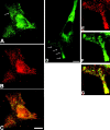
Figure 1.
Figure 1.
Distribution of β-actin mRNA and ZBP1 in CEFs. ZBP1 localizes in granular structures (arrow) that, along with β-actin mRNA, are enriched in cell lamellae. Immunofluorescence of ZBP1 (A) and in situ hybridization of β-actin mRNA (B) in serum-stimulated CEFs. (C) Overlap of A and C. CEFs were transiently transfected with GFP-tagged ZBP1 (D) and subsequently hybridized with β-actin mRNA fluorescent oligonucleotide (E). GFP–ZBP1 forms granules that are enriched at the cell periphery. Partial masking of granules is occurring due to high signal intensities. The enlargements (3×) (E–G) of indicated region in D (arrows) demonstrate a spatial overlap (G) of β-actin probe (E) and GFP–ZBP1 (F) within the cell protrusion. Bars, 10 μm.
Figure 2.
Figure 2.
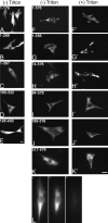
Cytoplasmic granule formation and cytoskeletal anchoring is mediated by KH3-KH4 of ZBP1. ZBP1 fragments fused to GFP were transiently transfected into CEFs. Localization of GFP-tagged proteins was analyzed before (A–J) and after (F'–J') extraction with Triton X-100. (A–E) Note that all protein fragments lacking the two COOH-terminal KH domains (KH3 and KH4) in their entirety were evenly distributed throughout the cytoplasm (A, 1–179; B, 1–289; C, 1–397; D, 195–522; E, 195–403) and no granular structures were observed. All of these fusion proteins were extracted from the cytoplasm by treatment with Triton X-100 before fixation (L). (F–K) CEFs transiently transfected with: F, 1–576; G, 1–556; H, 74–576; I, 84–576; J, 189–576; and K, 317–576. The NH2-terminal truncated fragments formed large granules that were resistant to detergent extraction. The left column (F–J) shows nonextracted fixed cells. Cells in the right column (F'–J') have been Triton extracted. Note that granules formed by constructs in G (1–556) and H (74–576) are enriched in the lamellae, whereas granules formed by the remaining constructs, I (84–576), J (189–576), and K (317–576), were evenly distributed in the cytoplasm. (L) To demonstrate the loss of COOH-terminal truncated GFP fusion proteins by extraction, the loss of GFP signal was visualized after the introduction of Triton X-100 (t0). The COOH-terminal truncated fragment (1–397) was initially diffusely distributed in the cytoplasm. No significant signal was detected after 20 s of Triton extraction. Images shown are at introduction of Triton X-100 (t0), 10 s post-Triton (t10), and 20 s post-Triton (t20). Bars, 10 μm.
Figure 3.
Figure 3.
Localization phenotypes of GFP–ZBP1 deletion mutants. Red bars indicate putative nuclear localization signals, green bars indicate putative nuclear export signals, and yellow bars indicate putative map kinase sites.
Figure 4.
Figure 4.
Anchoring of ZBP1 granules to the actin cytoskeleton is mediated by KH domains. CEF cells were transfected with GFP-tagged ZBP1 (A–C) or GFP-tagged KH1-KH4 (189–576) (D–F). After treatment with either cytochalasin D or colchicine, cells were Triton extracted, fixed, labeled, and viewed. Localization of GFP proteins on the microfilament (Phalloidin) or microtubule (anti-tubulin) systems was visualized and compared. In colchicine-treated cells, the microtubule system was significantly diminished; however, both GFP-tagged proteins were still associated with granular structures and retained their typical localization compared with untreated cells. In contrast, treatment with cytochalasin D destroyed the microfilament system and caused a significant loss of protein after extraction. Residual granular staining was only observed in the perinuclear region, indicating that granule formation and specific subcellular distribution mediated by the KH domains is mainly based on the actin cytoskeleton.
Figure 5.
Figure 5.
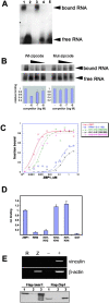
The KH domains of ZBP1 specifically bind the zipcode of β-actin mRNA. (A) Electrophoretic mobility shift assay of 32P-labeled zipcode RNA with recombinant GST–ZBP1 fragments. ZBP1 fragments were incubated with labeled zipcode in the presence of E. coli tRNA nonspecific competitor followed by heparin. (Lane 1) ZBP1; (lane 2) RRM1–2 (1–195); (lane 3) KH1-KH4 (195–576); (lane 4) GST; (lane 5) no protein. Note that only the full-length protein and the fragment containing the KH domains (lanes 1 and 3) caused a significant shift of the labeled RNA. No binding was observed for the RRM domains or GST (2 and 4). (B) Interaction between zipcode RNA and ZBP1 was reduced by addition of wild-type zipcode RNA but not by mutated zipcode RNA. (C) Nitrocellulose filter binding assay of recombinant GST–ZBP1 fragment affinity for β-actin mRNA zipcode: ZBP1 (1–576), RRM1-2 (1–195), KH1-KH4 (195–576), KH1-2 (195–308), KH3-KH4 (404–576). Recombinant proteins at various concentrations were incubated with 32P-labeled zipcode RNA. Bound probe was detected by Cerenkov counting after binding of the protein–RNA complex to the filter and intensive washing. All fragments containing at least KH domain 3 and 4 bound the zipcode with a K d in the nM range. (D) To determine the relative affinities of GST–ZBP1 fragments for full-length β-actin, mRNA binding was analyzed in GST pull-down assays. The fraction of bound 32P-labeled human β-actin RNA retained on equal amounts of immobilized GST–ZBP1 proteins were normalized to binding of full-length ZBP1 and plotted according to the fragments used. All proteins containing at least KH3 and 4 bound the full-length mRNA at apparently equal affinity. (E) Human β-actin mRNA coimmunoprecipitates specifically with Flag-tagged ZBP1. RT-PCR amplification of vinculin or β-actin mRNA extracted from supernatant of nontransfected cells (+) or pellets of FLAG–ZBP1 (Z), FLAG–raver1 (R), or mock (−)-transfected cells after immunoprecipitation with anti-Flag (M2). Human β-actin mRNA was specifically enriched in FLAG–ZBP1 pellets, whereas no enrichment of vinculin mRNA was detected. Precipitation of the FLAG-tagged raver1 (left, bottom) or ZBP1 (right, bottom) was verified by Western blotting of pellet fractions using anti-Flag (M2) antibody. (Lane 1) Total cell extract; (lane 2) supernatant after immunoprecipitation; (lane 3) pelleted fraction.
Figure 6.
Figure 6.
Selection of ZBP1 (KH3-KH4) ligands using RNA selection amplification (SELEX). (A) Lowercase letters are fixed sequences present in RNA library clones. Boxed sequences indicate consensus sequence. (B) Interaction of ZBP1 with zipcode RNA was reduced by addition of 20-mer oligonucleotides that bear ACACCC repeats of the zipcode or consensus SELEX sequences (S1).
Figure 7.
Figure 7.
ZBP1 plays a role in β-actin mRNA localization. CEFs were transfected with deletion mutants of ZBP1 fused to GFP. Constructs are schematically represented to the left of the table as follows: RRM domains are yellow with green hatching, KH domains are in black, putative nuclear export signal are in green, putative nuclear localization signals are in red, putative MAP kinase site are in yellow. After fixation and permeabilization, in situ hybridization was performed to detect β-actin mRNA. Bars show the percentage of transfected cells counted with β-actin mRNA localized to leading edge. At least 100 cells were counted blind per coverslip in three experiments each. Routinely, 40–50% of nontransfected CEFs show β-actin mRNA localization (unpublished data). (a, 1–179; b, 1–289; c, 1–397; d, 74–576; e, 84–576; f, 189–576; g, 317–576; h, 195–403; i, 195–522; j, 1–576 [ZBP1]).

References

    1. Barbarese, E., D.E. Koppel, M.P. Deutscher, C.L. Smith, K. Ainger, F. Morgan, and J.H. Carson. 1995. Protein translation components are colocalized in granules in oligodendrocytes. J. Cell Sci. 108:2781–2790. - PubMed
    1. Bashirullah, A., R.L. Cooperstock, and H.D. Lipshitz. 1998. RNA localization in development. Annu. Rev. Biochem. 67:335–394. - PubMed
    1. Bassell, G.J., H. Zhang, A.L. Byrd, A.M. Femino, R.H. Singer, K.L. Taneja, L.M. Lifshitz, I.M. Herman, and K.S. Kosik. 1998. Sorting of beta-actin mRNA and protein to neurites and growth cones in culture. J. Neurosci. 18:251–265. - PMC - PubMed
    1. Broadus, J., and C.Q. Doe. 1997. Extrinsic cues, intrinsic cues and microfilaments regulate asymmetric protein localization in Drosophila neuroblasts. Curr. Biol. 7:827–835. - PubMed
    1. Carey, J., V. Cameron, P.L. de Haseth, and O.C. Uhlenbeck. 1983. Sequence-specific interaction of R17 coat protein with its ribonucleic acid binding site. Biochemistry. 22:2601–2610. - PubMed

Publication types

MeSH terms