. 2003 Jan 6;160(1):89-99.
doi: 10.1083/jcb.200210115. Epub 2002 Dec 30.

Roles of G beta gamma in membrane recruitment and activation of p110 gamma/p101 phosphoinositide 3-kinase gamma

Affiliations

Roles of G beta gamma in membrane recruitment and activation of p110 gamma/p101 phosphoinositide 3-kinase gamma

Carsten Brock et al. J Cell Biol. .

Abstract

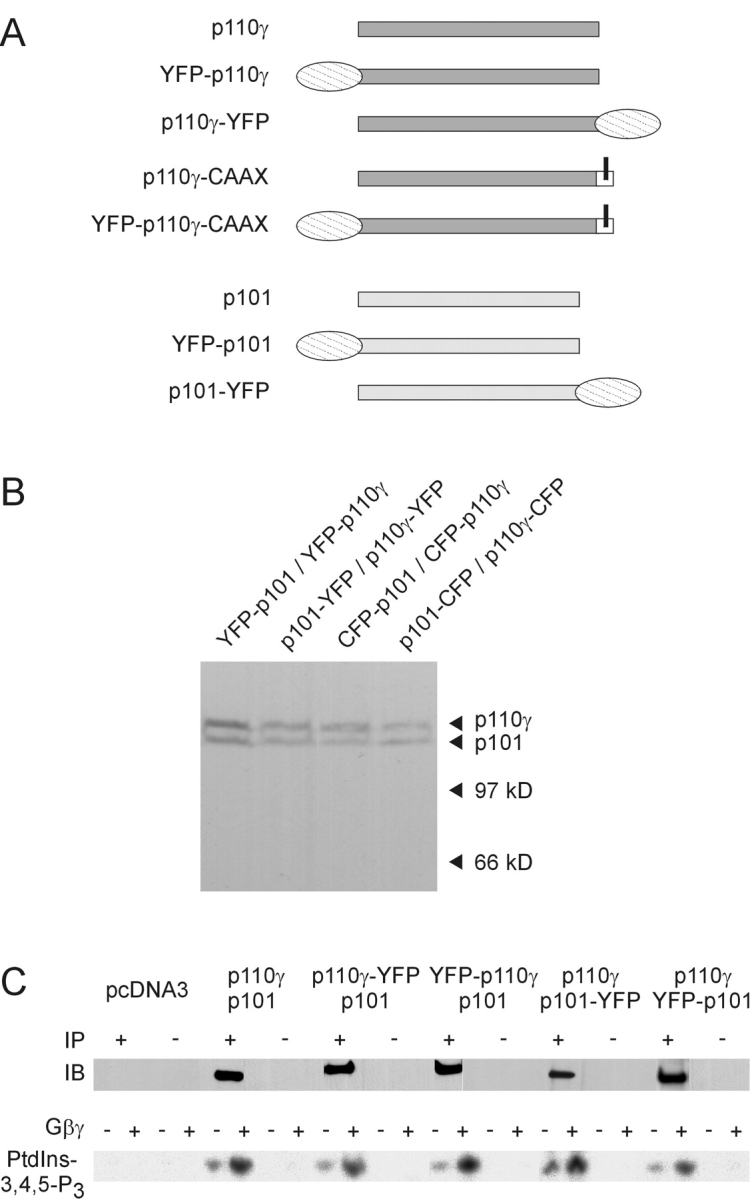
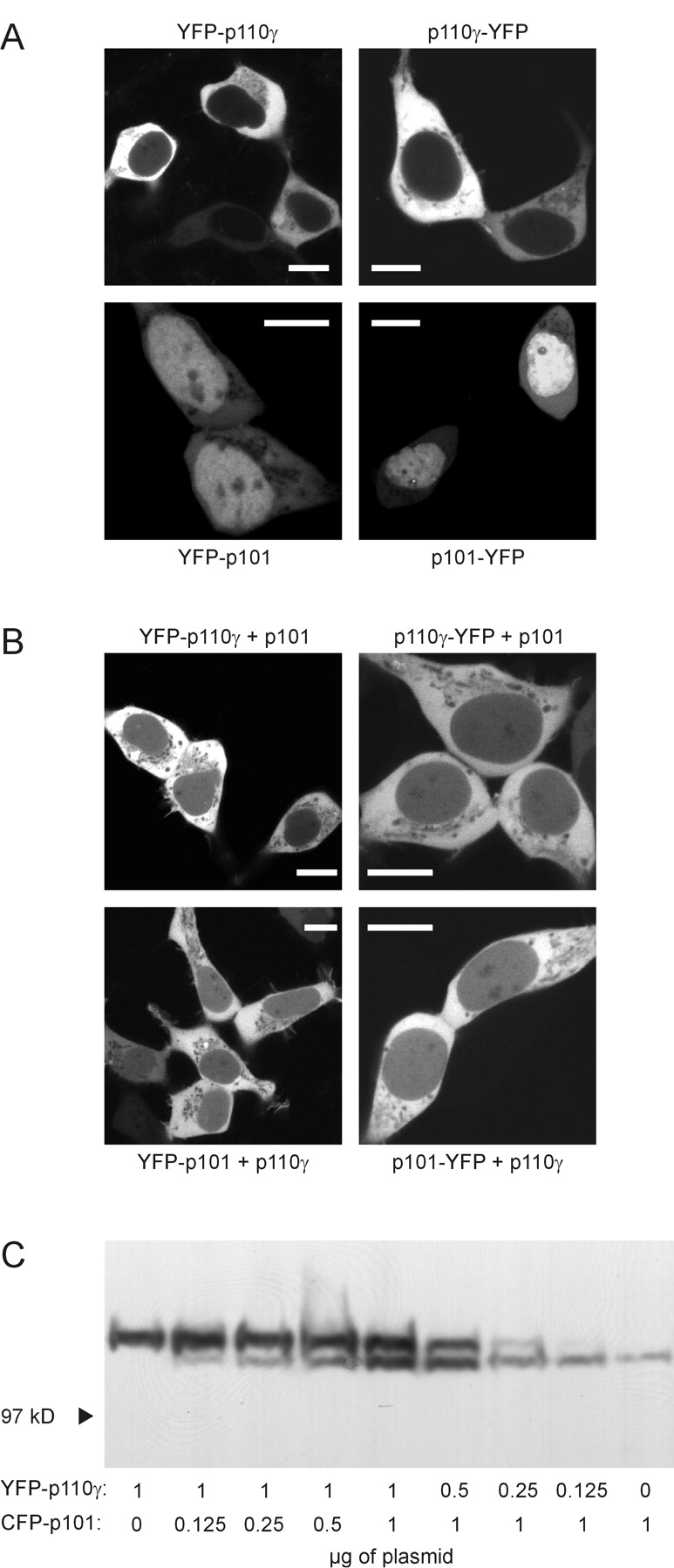
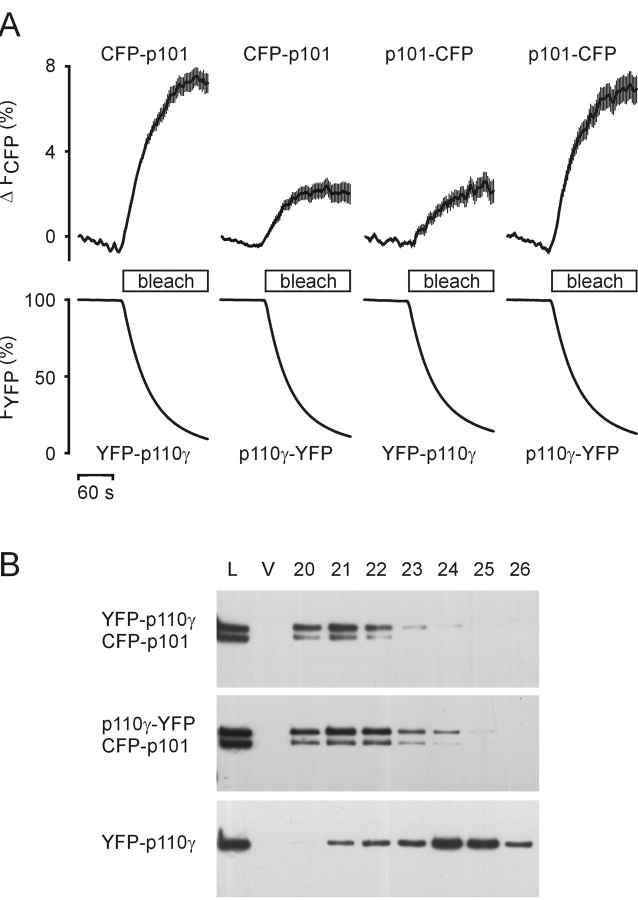
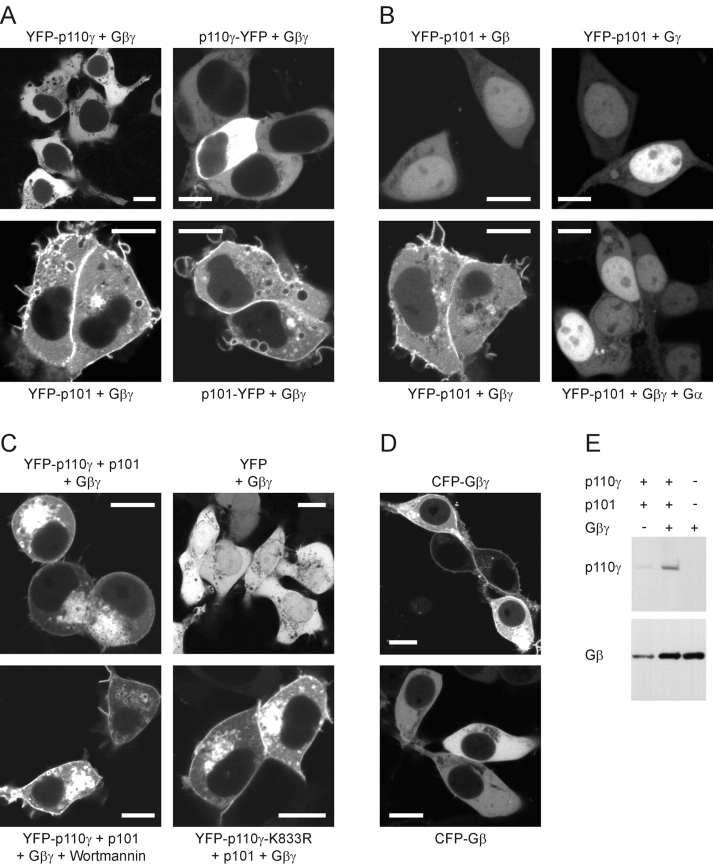
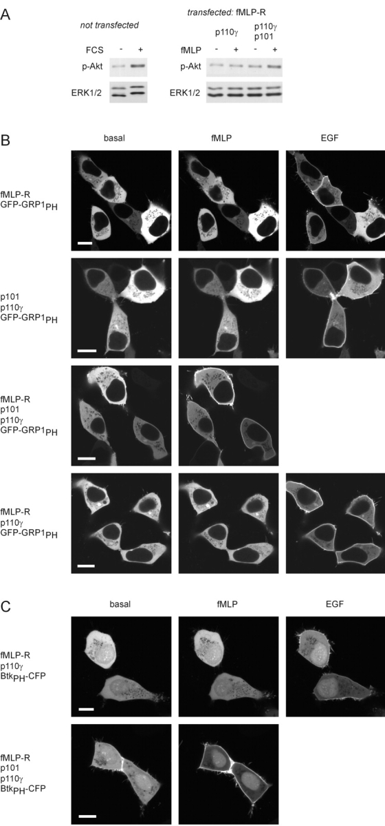
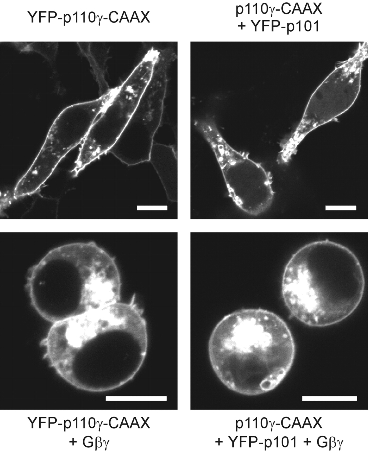
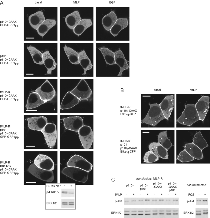
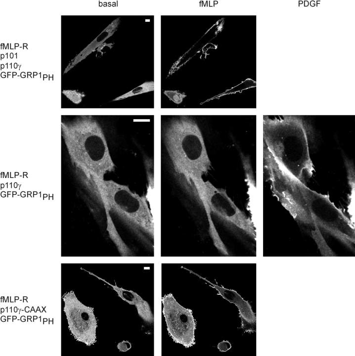
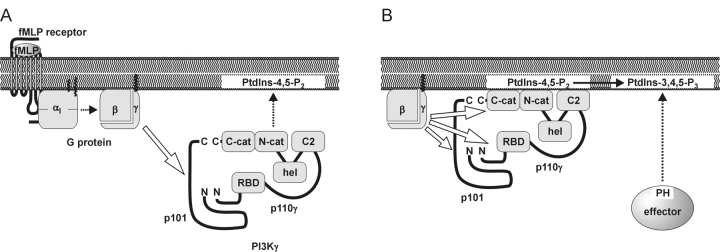
Receptor-regulated class I phosphoinositide 3-kinases (PI3K) phosphorylate the membrane lipid phosphatidylinositol (PtdIns)-4,5-P2 to PtdIns-3,4,5-P3. This, in turn, recruits and activates cytosolic effectors with PtdIns-3,4,5-P3-binding pleckstrin homology (PH) domains, thereby controlling important cellular functions such as proliferation, survival, or chemotaxis. The class IB p110 gamma/p101 PI3K gamma is activated by G beta gamma on stimulation of G protein-coupled receptors. It is currently unknown whether in living cells G beta gamma acts as a membrane anchor or an allosteric activator of PI3K gamma, and which role its noncatalytic p101 subunit plays in its activation by G beta gamma. Using GFP-tagged PI3K gamma subunits expressed in HEK cells, we show that G beta gamma recruits the enzyme from the cytosol to the membrane by interaction with its p101 subunit. Accordingly, p101 was found to be required for G protein-mediated activation of PI3K gamma in living cells, as assessed by use of GFP-tagged PtdIns-3,4,5-P3-binding PH domains. Furthermore, membrane-targeted p110 gamma displayed basal enzymatic activity, but was further stimulated by G beta gamma, even in the absence of p101. Therefore, we conclude that in vivo, G beta gamma activates PI3K gamma by a mechanism assigning specific roles for both PI3K gamma subunits, i.e., membrane recruitment is mediated via the noncatalytic p101 subunit, and direct stimulation of G beta gamma with p110 gamma contributes to activation of PI3K gamma.


Figures

Figure 1.
Figure 1.
Construction and characterization of fluorescent PI3Kγ fusion proteins. (A) Schematic representation of the wild-type, fluorescent, and membrane-targeted PI3Kγ subunits. A YFP (or CFP) tag was fused to either the NH2- or the COOH terminus of p110γ and p101. The p110γ-CAAX fusion protein contains an isoprenylation motif (bars for lipid modifications). (B) Differently fluorescence-tagged PI3Kγ subunits were coexpressed in HEK cells, and cell lysates were subjected to SDS-PAGE followed by immunoblotting with a GFP-specific antibody. (C) Catalytic activity of fluorescent PI3Kγ fusion proteins. HEK cells were transfected with the indicated plasmids. Lysates were subjected to immunoprecipitation (IP) with (+) or without (−) an anti-p110γ antibody. immunoprecipitation was controlled by immunoblotting (IB) with another anti-p110γ antibody. Shown are chemiluminescence images (top panels). Immunoprecipitates were assayed for in vitro PI3K activity in the absence or presence of 120 nM Gβγ using PtdIns-4,5-P2– containing lipid vesicles and γ[32P]ATP as substrates. Depicted are autoradiographs of the generated 32P-PtdIns-3,4,5-P3 (bottom panels).
Figure 2.
Figure 2.
Expression and subcellular distribution of fluorescent p110γ and p101 in HEK cells. (A) Cells were transfected with plasmids for YFP-tagged single PI3Kγ subunits as indicated. Images were taken by confocal laser scanning microscopy. Images of typical cells are shown. White bars indicate a 10-μm scale. (B) Cells were cotransfected with both p110γ and p101. Only one subunit was fused to YFP as indicated. (C) Cells were transfected with plasmids encoding fluorescent p110γ and p101 at different ratios. The total amount of transfected cDNA was kept constant by the addition of pcDNA3. Equal amounts of whole-cell lysates were subjected to SDS-PAGE followed by immunoblotting with an anti-GFP antibody.
Figure 3.
Figure 3.
Dimerization of p110γ and p101. (A) HEK cells were cotransfected with plasmids encoding NH2- or COOH-terminally YFP-tagged p110γ and NH2- or COOH-terminally CFP-tagged p101 in different combinations. FRET was measured in vivo. An increase in CFP (donor) fluorescence during YFP (acceptor) bleach indicates FRET between fluorescent PI3Kγ subunits. The depicted data represent means ± SEM of at least 18 single cells in three independent transfection experiments. (B) HEK cells were transfected with the indicated plasmids. Cytosols were prepared and subjected to gel filtration. The elution profiles were analyzed by immunoblotting with an anti-GFP antibody (L, load diluted 1:5; V, void volume; 20–26, fraction numbers).
Figure 4.
Figure 4.
Membrane recruitment of PI3Kγ by Gβγ. The effect of coexpression of Gβγ on the subcellular distribution of p110γ, p101, and heterodimeric PI3Kγ in HEK cells was analyzed by confocal laser scanning microscopy. Images of typical cells are shown (white bars, 10 μm). (A) Coexpression of monomeric PI3Kγ subunits with Gβγ. (B) Controls; coexpression of YFP-p101 with Gβ1, Gγ2, Gβ1γ2, or Gβ1γ2 and the Gβγ- scavenging Gαi2. (C) Coexpression of heterodimeric PI3Kγ with Gβγ. Note the round shape of the cells. Controls; coexpression of Gβγ with YFP or with kinase-deficient YFP-p110γ-K833R and p101; effect of 100 nM wortmannin. (D) Subcellular localization of Gβγ. Cells were transfected with a plasmid encoding CFP-tagged Gβ1 alone or together with the Gγ2 plasmid. (E) Immunoblot analysis of membrane fractions. HEK cells were transfected with the plasmids encoding PI3Kγ, Gβγ, or both together. To avoid rounding and detachment of the cells, the kinase-deficient mutant YFP-p110γ-K833R was used. Membrane fractions were prepared and analyzed by immunoblotting with anti-p110γ and anti-Gβ antibodies.
Figure 5.
Figure 5.
Activation of PI3Kγ by a G protein–coupled receptor. (A) Akt phosphorylation. Whole-cell lysates were analyzed by immunoblotting using an antibody that specifically recognizes the phosphorylated form of Akt. Equal loading was shown by using an anti-ERK antibody. Left; FCS-induced Akt phosphorylation in untransfected HEK cells. Right; fMLP-induced Akt phosphorylation in cells expressing the fMLP receptor and only the catalytic (p110γ) or both PI3Kγ subunits. (B) Membrane recruitment of the PtdIns-3,4,5-P3–binding PH domain of GRP1. HEK 293 cells were transfected with plasmids encoding the PH domain of GRP1 fused to GFP (GFP-GRP1PH), and the human fMLP receptor (fMLP-R), p110γ, and p101 in different combinations. The localization of the GFP-GRP1PH was monitored before and after the addition of 1 μM fMLP and 100 ng/ml EGF (added 8 min later) by confocal laser scanning microscopy. Pictures were taken 4 min after addition of either agonist. Images of typical experiments are shown (white bars, 10 μm). (C) Membrane recruitment of the PtdIns-3,4,5-P3–binding PH domain of Btk. Instead of GFP-GRP1PH, the PH domain of Btk fused to CFP (BtkPH-CFP) was used as a PtdIns-3,4,5-P3 sensor.
Figure 6.
Figure 6.
Characterization of a constitutively membrane-associated p110γ-CAAX. HEK cells were transfected with the indicated plasmids and analyzed by confocal laser scanning microscopy. Images of typical cells are shown. White bars indicate a 10-μm scale. Top panel; Subcellular localization of YFP-p110γ-CAAX (left) or YFP-p101 expressed together with p110γ-CAAX (right). Bottom panel; Coexpression with Gβγ.
Figure 7.
Figure 7.
Activation of a constitutively membrane-associated p110γ-CAAX. (A) GFP-GRP1PH translocation. HEK cells were transfected with plasmids for GFP-GRP1PH and p110γ-CAAX, p101, fMLP-R, and dominant-negative Ras N17 in different combinations. Translocation of the PtdIns-3,4,5-P3–binding GFP-GRP1PH in response to agonist stimulation was monitored as described above (see Fig. 5). White bars indicate a 10-μm scale. As a positive control for H-Ras N17, cells were transfected with the H-Ras N17 plasmid (same amount as for the GFP-GRP1PH translocation experiment) or empty vector. Cells were stimulated with 10 ng/ml EGF, and whole-cell lysates were analyzed by immunoblotting with an antibody that specifically recognizes the phosphorylated form of ERK (p-ERK). Equal loading was shown using the anti-ERK-antibody. (B) BtkPH-CFP translocation. (C) Akt phosphorylation. The experiment shown in Fig. 5 was repeated. In addition, cells were transfected with the plasmid for the membrane-targeted p110γ-CAAX instead of wild-type p110γ.
Figure 8.
Figure 8.
PI3Kγ-mediated membrane translocation of GFP-GRP1 PH in vascular smooth muscle (VSM) cells. VSM cells were microinjected with plasmids for GFP-GRP1PH, fMLP-R, p101, and p110γ or p110γ-CAAX in different combinations. The localization of GFP-GRP1PH was monitored before and after the addition of 1 μM fMLP (picture taken after 4 min) and 10 ng/ml PDGF-BB (added 12 min later, picture taken after 8 min). White bars indicate a 10-μm scale.
Figure 9.
Figure 9.
Hypothetical model for receptor- induced membrane recruitment and activation of heterodimeric PI3Kγ. fMLP receptor, a prototypical heptahelical receptor coupled to Gi proteins. PI3Kγ; the cytosolic enzyme consists of a noncatalytic p101 subunit, which is in a tight complex with p110γ, thereby stabilizing p101. The NH2 and COOH termini of p101 and p110γ are oriented in close proximity, respectively. Contact sites involve the NH2 termini of both subunits. The modular domain structure of p110γ (RBD, Ras binding domain; C2, C2 domain; hel, helical domain; N-cat, C-cat, NH2- and COOH-terminal lobes of the catalytic domain) is based on the crystal structure of an NH2-terminally truncated p110γ. (A) Membrane recruitment. The agonist-stimulated receptor induces the release of Gβγ from Gi proteins (dotted arrow). Gβγ recruits the PI3Kγ heterodimer to the plasma membrane (dotted arrow) by binding to the noncatalytic p101 (hollow arrow). Accordingly, Gβγ and p101 function as a membrane anchor and an adaptor for PI3Kγ, respectively. In addition, the C2 domain of p110γ may facilitate membrane attachment through interaction with phospholipids. p101 may also affect the interaction of PI3Kγ with the lipid interface. Membrane-attached PI3Kγ exhibits basal enzymatic activity. (B) Allosteric activation. At the membrane, Gβγ activates PI3Kγ by direct interaction with p110γ (hollow arrows). This stimulation does not require p101. However, p101 may participate in Gβγ-induced stimulation of membrane-attached p110γ. The stoichiometry of the Gβγ–PI3Kγ interaction is unknown, i.e., Gβγ may interact with p101 and p110γ through individual or common binding sites. Gβγ binds to the NH2- and COOH-terminal part of p110γ, the latter harboring the catalytic domain. Thus, Gβγ may allosterically activate PI3Kγ through a conformational change in the catalytic domain. Accordingly, Gβγ significantly increases Vmax of PtdIns-3,4,5-P3 production. PtdIns-3,4,5-P3, in turn, recruits PH domain–containing effectors such as GRP1 or Btk to the plasma membrane (dotted arrow).

References

    1. Al-Aoukaty, A., B. Rolstad, and A.A. Maghazachi. 1999. Recruitment of pleckstrin and phosphoinositide 3-kinase γ into the cell membranes, and their association with Gβγ after activation of NK cells with chemokines. J. Immunol. 162:3249–3255. - PubMed
    1. Balla, T., T. Bondeva, and P. Várnai. 2000. How accurately can we image inositol lipids in living cells? Trends Pharmacol. Sci. 21:238–241. - PubMed
    1. Baier, R., T. Bondeva, R. Klinger, A. Bondev, and R. Wetzker. 1999. Retinoic acid induces selective expression of phosphoinositide 3-kinase γ in myelomonocytic U937 cells. Cell Growth Differ. 10:447–456. - PubMed
    1. Bondev, A., T. Bondeva, and R. Wetzker. 2001. Regulation of phosphoinositide 3-kinase γ protein kinase activity in vitro and in COS-7 cells. Signal Transduction. 1:79–85.
    1. Bondeva, T., L. Pirola, G. Bulgarelli-Leva, I. Rubio, R. Wetzker, and M.P. Wymann. 1998. Bifurcation of lipid and protein kinase signals of PI3Kγ to the protein kinases PKB and MAPK. Science. 282:293–296. - PubMed

Publication types

MeSH terms

Substances