

Angiostatin up-regulation in gastric cancer cell SGC7901 inhibits tumorigenesis in nude mice
- PMID: 12508352
- PMCID: PMC4728250
- DOI: 10.3748/wjg.v9.i1.59
Angiostatin up-regulation in gastric cancer cell SGC7901 inhibits tumorigenesis in nude mice
Abstract
Aim: To explore the influence of angiostatin up-regulation on the biologic behavior of gastric cancer cells in vitro and in vivo, and the potential of angiostatin gene therapy in the treatment of human gastric cancer.
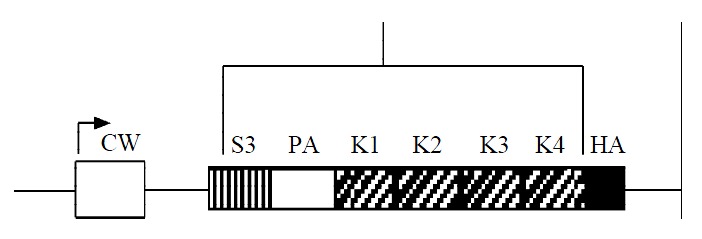
Methods: Mouse angiostatin cDNA was subcloned into the eukaryotic expression vector pcDNA3.1(+) and identified by restriction endonucleases digestion and sequencing. The recombinant vector pcDNA3.1(+)-angio was transfected into human gastric cancer cells SGC7901 with liposome and paralleled with the vector control and the mock control. Angiostatin transcription and protein expression were examined by RT-PCR and Western blot in the stable cell lines selected by G418. Cell proliferation and growth in vitro of the three groups were observed respectively under microscope, cell number counting and FACS. The cells overexpressing angiostatin, vector transfected and untreated were respectively implanted subcutaneously into nude mice. After 30 days the size of tumors formed was measured, and microvessel density count (MVD) in the tumor tissues was assessed by immunohistochemistry with the primary anti-vWF antibody.
Results: The recombinant vector pcDNA3.1(+)-angio was confirmed with the correct sequence of mouse angiostatin under the promoter CMV. After 30 d of transfection and selection with G418, macroscopic resistant cell clones were formed in the experimental group transfected with pcDNA 3.1(+)-angio and the vector control. But no untreated cells survived in the mock control. Angiostatin mRNA transcription and protein expression were detected in the experimental group. No significant differences were observed among the three groups in cell morphology, cell growth curves and cell cycle phase distributions in vitro. However, in nude mice model, markedly inhibited tumorigenesis and slowed tumor expansion were observed in the experimental group as compared with the controls, which was paralleled with decreased microvessel density in and around tumor tissues (P<0.05).
Conclusion: Angiostatin does not directly inhibit human gastric cancer cell proliferation and growth in vitro, but exerts its anti-tumor functions through antiangiogenesis in a paracrine way in vivo.
Figures


References
-
- Rosen LS. Angiogenesis inhibition in solid tumors. Cancer J. 2001;7 Suppl 3:S120–S128. - PubMed
-
- Sugimachi K, Tanaka S, Terashi T, Taguchi K, Rikimaru T, Sugimachi K. The mechanisms of angiogenesis in hepatocellular carcinoma: angiogenic switch during tumor progression. Surgery. 2002;131:S135–S141. - PubMed
-
- Cao Y. Endogenous angiogenesis inhibitors and their therapeutic implications. Int J Biochem Cell Biol. 2001;33:357–369. - PubMed
-
- Hagedorn M, Bikfalvi A. Target molecules for anti-angiogenic therapy: from basic research to clinical trials. Crit Rev Oncol Hematol. 2000;34:89–110. - PubMed
Publication types
MeSH terms
Substances
LinkOut - more resources
Full Text Sources
Medical
Miscellaneous
