. 2003 Jan;23(2):482-92.
doi: 10.1128/MCB.23.2.482-492.2003.

Alteration of mesodermal cell differentiation by EWS/FLI-1, the oncogene implicated in Ewing's sarcoma

Affiliations

Alteration of mesodermal cell differentiation by EWS/FLI-1, the oncogene implicated in Ewing's sarcoma

Susan Eliazer et al. Mol Cell Biol. 2003 Jan.

Abstract

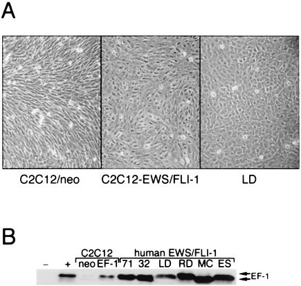
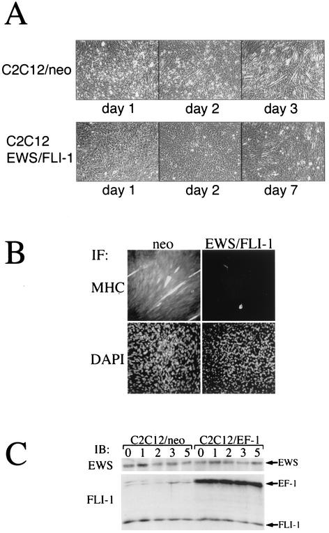
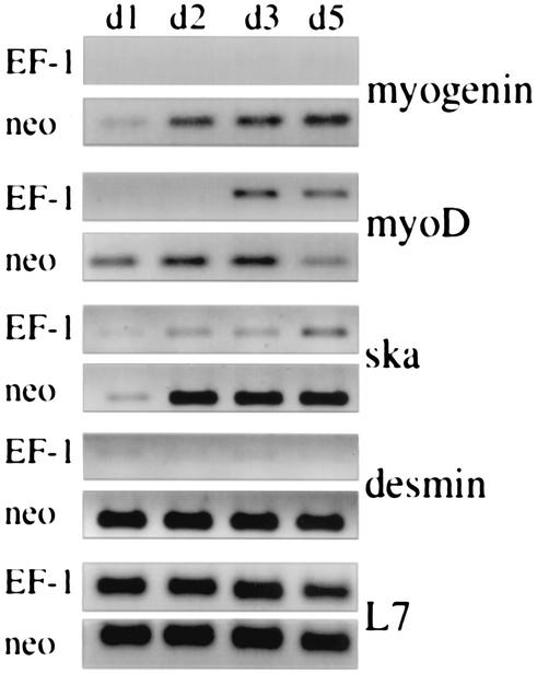
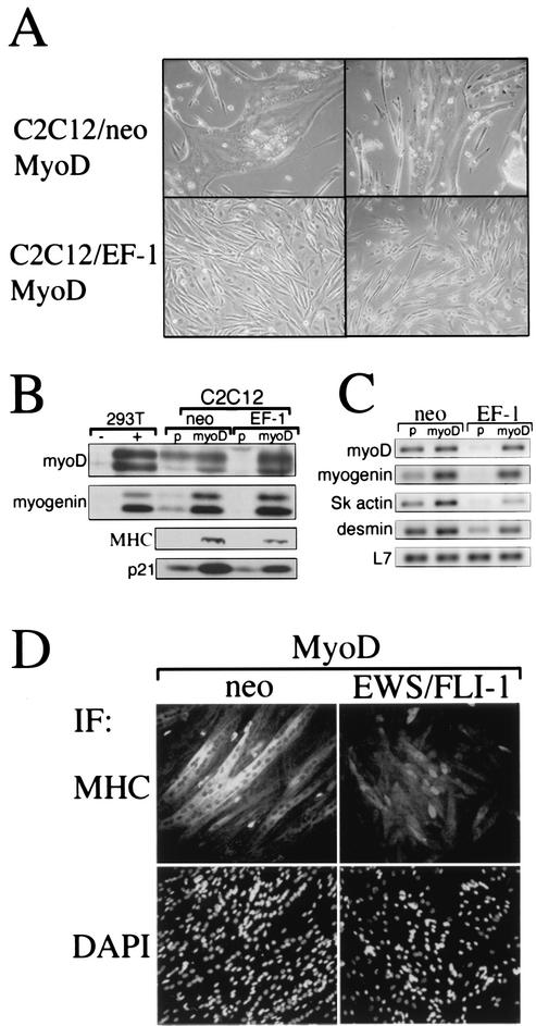
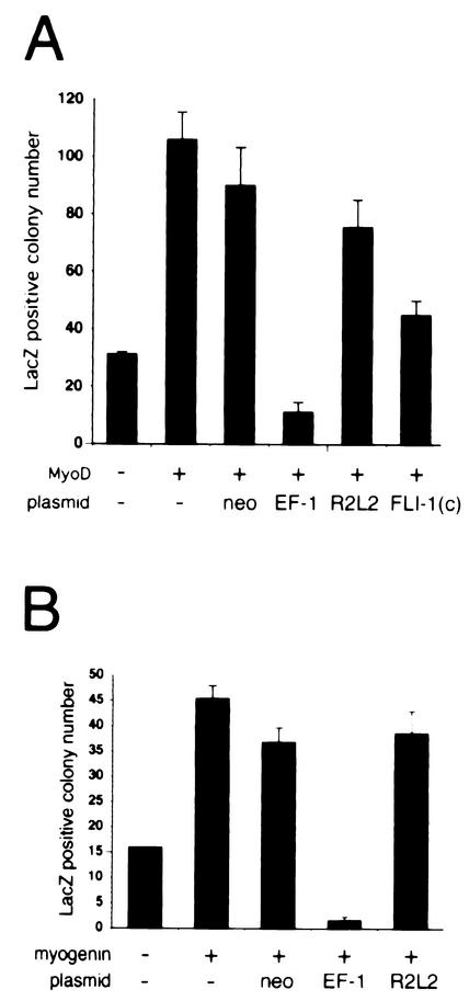
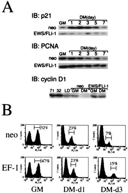
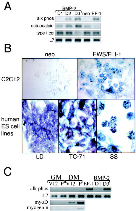
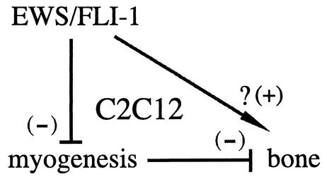
The chimeric fusion gene EWS/FLI-1 is detected in most cases of Ewing's sarcoma (ES), the second most common malignant bone tumor of childhood. Although 80% of ES tumors develop in skeletal sites, the remainder can arise in almost any soft tissue location. The lineage of the cell developing the EWS/FLI-1 gene fusion has not been fully characterized but is generally considered to be of either mesenchymal or neural crest origin. To study this oncogene in a conceptually relevant target cell, EWS/FLI-1 was introduced into the murine cell line C2C12, a myoblast cell line capable of differentiation into muscle, bone, or fat. In this cellular context, EWS/FLI-1 profoundly inhibited the myogenic differentiation program. The block in C2C12 myogenic differentiation required the nuclear localization and DNA-binding functions of EWS/FLI-1 and was mediated by transcriptional and posttranscriptional suppression of the myogenic transcription factors MyoD and myogenin. Interestingly, C2C12-EWS/FLI-1 cells constitutively expressed alkaline phosphatase, a bone lineage marker, and were alkaline phosphatase positive by histochemistry but showed no other evidence of bone lineage commitment. Consistent with recent findings in human ES tumor cell lines, C2C12-EWS/FLI-1 cells constitutively expressed cyclin D1 and demonstrated decreased expression of the cell cycle regulator p21(cip1), even under differentiation conditions and at confluent density. This C2C12-EWS/FLI-1 cell model may assist in the identification of novel differentially expressed genes relevant to ES and provide further insight into the cell(s) of origin developing ES-associated genetic fusions.


Figures

FIG. 1.
FIG. 1.
Expression of EWS/FLI-1 in the multipotential cell line C2C12. (A) A stable polyclonal population of C2C12 cells expressing either type I EWS/FLI-1 (left) or vector alone (neo) (right) is shown at confluence in standard GM. Note the cuboidal appearance of the C2C12-EWS/FLI-1 cells compared to the more-spindle-shaped control C2C12 cells. For comparison, a human EWS/FLI-1 cell line (LD) is shown at the far right. (B) C2C12-EWS/FLI-1, C2C12-neo, and the human ES cell lines TC-71 (71), TC-32 (32), LD, RD-ES (RD), SK-N-MC (MC), and SK-ES-1 (ES) were analyzed by anti-FLI-1 immunoblotting.
FIG. 2.
FIG. 2.
EWS/FLI-1 inhibits the morphological differentiation of C2C12 cells to muscle. (A) C2C12-neo (top) or C2C12-EWS/FLI-1 cells were placed in DM (2% horse serum) for the indicated number of days and evaluated for morphological evidence of myogenic differentiation. Representative areas were then photographed. C2C12-neo cells differentiated to classic multinucleated myotubes by day 3 (top right), whereas C2C12-EWS/FLI-1 cells showed little evidence of morphological differentiation into myotubes, even after 7 days (bottom right) in DM. (B) C2C12-neo (neo) and C2C12-EWS/FLI-1 cells were analyzed on day 3 in DM by immunofluorescence (IF) with an antibody against MHC, a marker of myogenic differentiation, and stained with DAPI to visualize total nuclei. Note the only rare MHC-positive cell in the C2C12-EWS/FLI-1 population. (C) To confirm stable EWS/FLI-1 expression under differentiation conditions, C2C12-neo (neo) and C2C12-EWS/FLI-1 cells were analyzed by Western immunoblotting (IB) in GM (0) or after the indicated number of days in DM with an anti-FLI-1 antibody which recognizes endogenous FLI-1 and the EWS/FLI-1 fusion (EF-1) or with an antibody recognizing the endogenous EWS protein.
FIG. 3.
FIG. 3.
EWS/FLI-1 interferes with the induction of muscle-specific genes. C2C12 cells expressing EWS/FLI-1 (EF-1) or control vector (neo) were placed in DM, and the expression of myogenin, MyoD, skeletal actin (ska), and desmin was analyzed by semiquantitative RT-PCR (50) prepared on the indicated days (d1, d2, d3, and d5). To control for equivalent sample integrity and quantity, the expression of the ribosome-associated protein L7 mRNA is shown at the bottom. The data depicted are representative of three independent experiments.
FIG. 4.
FIG. 4.
Overexpression of MyoD fails to rescue C2C12-EWS/FLI-1 myogenic differentiation despite partial rescue of downstream gene expression. (A) C2C12-neo or C2C12-EWS/FLI-1 cells were transduced with a MyoD retrovirus and examined for morphological evidence of myogenic differentiation. Note that control cells transduced with MyoD retrovirus formed large multinucleated myotubes (upper photos) while EWS/FLI-1-expressing cells showed no evidence of myotube formation (lower photos). (B) Protein lysates from C2C12-neo or C2C12-EWS/FLI-1 cells were transduced with MyoD or control (p) retrovirus and examined by anti-MyoD, anti-myogenin, anti-MHC, or anti-p21 immunoblotting. As controls, 293T cells transfected with MyoD or myogenin are shown at the left (note that both myogenin and MyoD are detected as doublet bands). (C) The same cells depicted in panel B were analyzed by semiquantitative RT-PCR for myoD, myogenin, skeletal actin (sk actin), and desmin mRNA expression. The expression of the ribosome-associated protein L7 mRNA is shown at the bottom to confirm equivalent sample integrity. (D) C2C12-neo or C2C12-EWS/FLI-1 cells overexpressing MyoD were analyzed by immunofluorescence (IF) with an anti-MHC antibody. Note the less intense MHC staining of C2C12-EWS/FLI-1 cells compared to control (neo) cells. Cells were counterstained with DAPI (bottom) to visualize total cell nuclei.
FIG. 5.
FIG. 5.
EWS/FLI-1 blocks MyoD-dependent and myogenin-dependent transcriptional activation of a myogenin-lacZ reporter. 293T cells were transfected with MyoD (A) or myogenin (B) plasmid and a β-galactosidase (lacZ) reporter driven by 5′ regulatory regions from the myogenin gene (6, 7) and either control vector (neo), type I EWS/FLI-1 (EF-1), the point mutant EWS/FLI-1 R2L2, or a plasmid containing only the portion of FLI-1 present in the EWS/FLI-1 fusion [Fli-1(C)]. Transcriptional activation of the reporter was scored by the lacZ-positive colony number. Data were normalized for differences in transfection efficiency by cotransfection of a green fluorescent protein plasmid. The baseline myogenin-lacZ colony number (control plasmid and myogenin-lacZ only) is shown at the far left. Error bars represent the standard errors of duplicate experiments.
FIG. 6.
FIG. 6.
EWS/FLI-1 alters C2C12 cell cycle gene expression without interfering with DM-induced cell cycle arrest. Protein lysates were prepared from C2C12 cells expressing EWS/FLI-1 or control cells (neo) cultured in either GM or DM and analyzed by anti-p21 (upper panel), anti-PCNA (middle panel), or anti-cyclin D1 (lower panel) immunoblotting (IB). In the case of the cyclin D1, cells were analyzed in GM and after 3 days in DM. For comparison, protein lysates from the human ES cell lines TC-71, TC-32, and LD were probed with anti-cyclin D1 antibody and shown at the left. (B) C2C12-neo or C2C12-EWS/FLI-1 cells were cultured in GM or cultured for the indicated number of days in DM, labeled with BrdU, and then analyzed in an anti-BrdU-based immunofluorescence assay. The number of cells undergoing DNA replication (BrdU-positive cells) was quantitated by FACS, with the percentage of BrdU-positive cells shown on the right.
FIG. 7.
FIG. 7.
Signaling pathways important in muscle differentiation are intact in C2C12-EWS/FLI-1 cells. Protein lysates were prepared from C2C12-EWS/FLI-1 or vector control cells (neo) while in GM (day 0) or after the indicated number of days in muscle DM. Samples were analyzed by immunoblotting with an antibody recognizing the activated form of ERK1/2 (pERK) or total ERK1/2 (upper panel), an antibody recognizing the activated form of Akt1 (pAkt) or total Akt1/2 (middle panel), or an antibody recognizing the activated form of p38 MAPK (pp38) and total p38 (lower panel). Controls, consisting of NIH 3T3 cells starved (−) and stimulated (+) with serum (for ERK and Akt) and HeLa cells untreated (−) and treated (+) with anisomycin (for p38 MAPK), are shown at the left.
FIG. 8.
FIG. 8.
Constitutive alkaline phosphatase expression and activity in cells expressing EWS/FLI-1. (A) C2C12 cells expressing EWS/FLI-1 (EF-1) or vector control (neo) were analyzed for alkaline phosphatase (alk phos) expression by semiquantitative RT-PCR (top) while in GM. As a control for alkaline phosphatase expression, C2C12 cells were treated with BMP-2 and analyzed at the indicated day (D) number. To control for equivalent sample processing, the expression of the ribosome-associated protein L7 mRNA is shown at the bottom. (B) C2C12 cells expressing EWS/FLI-1 or control (neo) (top panel) and the human ES tumor cell lines LD, TC-71, and SS (bottom panel) were analyzed for alkaline phosphatase activity by alkaline phosphatase enzyme-based histochemistry. (C) C2C12 cells stably expressing V12 ras (V12) or the parental vector (P) were cultured in GM or for 3 days in DM and analyzed for the expression of alkaline phosphatase, MyoD, or myogenin by semiquantitative RT-PCR. As a control for alkaline phosphatase expression, C2C12 cells treated with BMP-2 are shown at the right. For comparison, the constitutive expression of alkaline phosphatase by C2C12-EWS/FLI-1 cells is also shown at the right. The expression of L7 is included as a control for sample integrity.
FIG. 9.
FIG. 9.
A working model of the effect of EWS/FLI-1 on C2C12 mesodermal differentiation. A schematic representation of EWS/FLI-1 inhibiting C2C12 myogenic signals, which normally act to suppress bone differentiation. The possibility that EWS/FLI-1 might also directly modulate bone lineage features is also depicted; see the text for additional details.

Similar articles

Cited by

References

    1. Arvand, A., and C. T. Denny. 2001. Biology of EWS/ETS fusions in Ewing's family tumors. Oncogene 20:5747-5754. - PubMed
    1. Bailly, R. A., R. Bosselut, J. Zucman, F. Cormier, O. Delattre, M. Roussel, G. Thomas, and J. Ghysdael. 1994. DNA-binding and transcriptional activation properties of the EWS-FLI-1 fusion protein resulting from the t(11;22) translocation in Ewing sarcoma. Mol. Cell. Biol. 14:3230-3241. - PMC - PubMed
    1. Bennett, A. M., and N. K. Tonks. 1997. Regulation of distinct stages of skeletal muscle differentiation by mitogen-activated protein kinases. Science 278:1288-1291. - PubMed
    1. Braun, T., E. Bober, G. Buschhausen-Denker, S. Kohtz, K. H. Grzeschik, H. H. Arnold, and S. Kotz. 1989. Differential expression of myogenic determination genes in muscle cells: possible autoactivation by the Myf gene products. EMBO J. 8:3617-3625. - PMC - PubMed
    1. Buckingham, M. 2001. Skeletal muscle formation in vertebrates. Curr. Opin. Genet. Dev. 11:440-448. - PubMed

Publication types

MeSH terms

LinkOut - more resources