. 2003 Jan;23(2):493-509.
doi: 10.1128/MCB.23.2.493-509.2003.

The 32-kilodalton subunit of replication protein A interacts with menin, the product of the MEN1 tumor suppressor gene

Affiliations

The 32-kilodalton subunit of replication protein A interacts with menin, the product of the MEN1 tumor suppressor gene

Karen E Sukhodolets et al. Mol Cell Biol. 2003 Jan.

Abstract

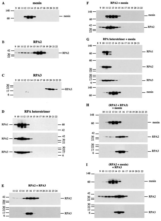
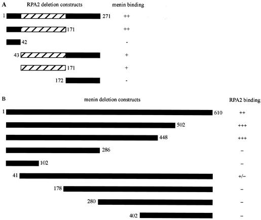
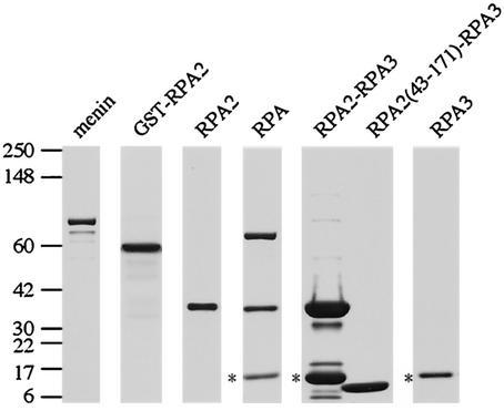
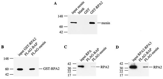
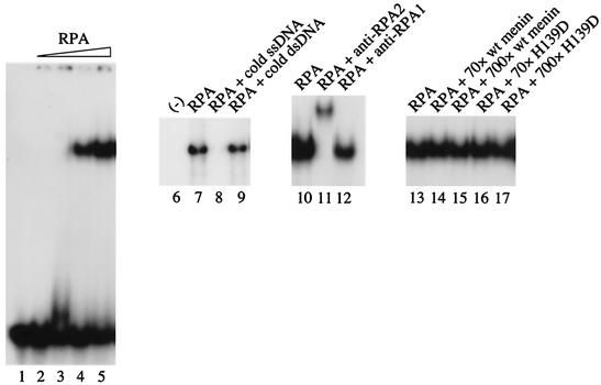
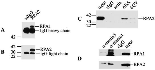
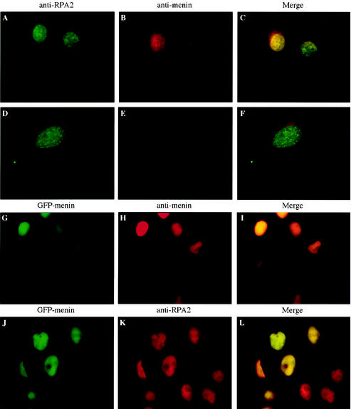
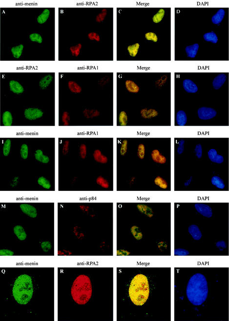
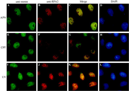
Menin is a 70-kDa protein encoded by MEN1, the tumor suppressor gene disrupted in multiple endocrine neoplasia type 1. In a yeast two-hybrid system based on reconstitution of Ras signaling, menin was found to interact with the 32-kDa subunit (RPA2) of replication protein A (RPA), a heterotrimeric protein required for DNA replication, recombination, and repair. The menin-RPA2 interaction was confirmed in a conventional yeast two-hybrid system and by direct interaction between purified proteins. Menin-RPA2 binding was inhibited by a number of menin missense mutations found in individuals with multiple endocrine neoplasia type 1, and the interacting regions were mapped to the N-terminal portion of menin and amino acids 43 to 171 of RPA2. This region of RPA2 contains a weak single-stranded DNA-binding domain, but menin had no detectable effect on RPA-DNA binding in vitro. Menin bound preferentially in vitro to free RPA2 rather than the RPA heterotrimer or a subcomplex consisting of RPA2 bound to the 14-kDa subunit (RPA3). However, the 70-kDa subunit (RPA1) was coprecipitated from HeLa cell extracts along with RPA2 by menin-specific antibodies, suggesting that menin binds to the RPA heterotrimer or a novel RPA1-RPA2-containing complex in vivo. This finding was consistent with the extensive overlap in the nuclear localization patterns of endogenous menin, RPA2, and RPA1 observed by immunofluorescence.


Figures

FIG.4.
FIG.4.
Menin binds preferentially to free RPA2 in gel filtration experiments with purified proteins. Binding reactions were performed by mixing purified proteins in approximately equimolar ratios as indicated and incubating them on ice for 30 min. The binding reaction mixtures were then injected onto an analytical gel filtration column, 0.2-ml fractions were collected, and portions of these fractions were analyzed by SDS-PAGE on 4 to 20% gradient minigels, followed by Western blotting with antibodies that recognize menin (SQV), RPA1 (αSSB70C), RPA2 (αSSB34A), or RPA3 (RPA14 11.1). The positions of the protein standards and their corresponding molecular masses (in kilodaltons) are indicated to the left or right of each panel. The migration of individual menin, RPA2, and RPA3 proteins is shown in panels A to C, respectively, and the migration of the RPA heterotrimer is shown in panel group D. The migration of menin mixed with RPA2 or the RPA heterotrimer is shown in panel groups F and G, respectively. The effect of menin on a preformed RPA2-RPA3 complex and of RPA3 on a preformed menin-RPA2 complex is shown in panel groups H and I, respectively, in comparison to a mixture of RPA2 and RPA3 (group E).
FIG. 1.
FIG. 1.
Mapping of regions involved in menin-RPA2 binding. Gal4 activation domain-RPA2 (A) and GAL4 DNA-binding domain-menin (B) fusion proteins containing the indicated regions (represented by black or hatched bars) were tested for interaction as described in Table 2, footnote a. The numbers to the left and right of the bars indicate the starting and ending amino acids, respectively, of the various menin and RPA2 constructs. The hatched area of RPA2 (amino acids 43 to 171) represents the region containing the ssDNA-binding domain.
FIG.2.
FIG.2.
Purified menin and RPA proteins used for in vitro binding studies. Purified proteins used in the experiments shown in Fig. 3 to 5 were analyzed by SDS-4 to 20% PAGE and stained with Coomassie blue. The positions of the protein standards and their corresponding molecular masses (in kilodaltons) are indicated on the left. Note that the RPA3 subunits present in the RPA heterotrimer and RPA2-RPA3 complex retained their His tags (indicated by asterisks) and therefore had a slightly reduced mobility compared to the untagged RPA3 subunit present in the truncated RPA2(43-171)-RPA3 complex. Also, the truncated RPA2(43-171) polypeptide and the RPA3 subunit were too similar in size to be resolved under these conditions, resulting in their appearance as a single band. However, the presence of both truncated RPA2 and RPA3 in the RPA(43-171)-RPA3 complex was confirmed by Western blotting (data not shown).
FIG.3.
FIG.3.
Menin binds preferentially to free RPA2 in pulldown experiments with purified proteins. Binding reactions were performed by mixing GST or GST-RPA2 bound to glutathione-Sepharose with purified menin (A) or by mixing FLAG-BAP or FLAG-menin bound to anti-Flag-agarose with various purified forms of RPA2, i.e., GST-RPA2 (B), the RPA heterotrimer (C), and the RPA2-RPA3 complex (D). After incubation for 1 h at 37°C, the beads were pelleted by brief microcentrifugation and washed several times to remove nonspecifically bound proteins. Bound proteins were eluted with protein sample buffer and heating for 10 min at 70°C and analyzed by SDS-4 to 20% PAGE, followed by Western blotting with antibodies that recognize menin (SQV) or RPA2 (αSSB34A). The positions of the protein standards are indicated to the left of each panel with their corresponding molecular masses (in kilodaltons).
FIG. 5.
FIG. 5.
Ability of RPA to bind ssDNA in vitro is not specifically influenced by menin. Binding reactions were performed by mixing purified wild-type or mutant menin (H139D) with RPA as indicated, followed by the addition of 32P-labeled (dC)30 and incubation for 20 min at room temperature. With the exception of the sample in lane 1, GST was also added to all of the binding reaction mixtures as a negative control/nonspecific stabilizing protein. Loading dye was then added, and the samples were analyzed by electrophoresis at 150 V on an 8% polyacrylamide gel in TBE (45 mM Tris-Cl, 45 mM boric acid, 1 mM EDTA) at room temperature, followed by vacuum drying at 70°C and autoradiography with an intensifying screen at −80°C. The components of the binding reactions were as follows: 25 fmol of (dC)30 (all lanes), ≈0.3 (lane 3), 3 (lane 4), or 30 (lanes 5 and 7 to 17) fmol of RPA, 2.5 pmol of unlabeled ssDNA (lane 8), 2 pmol of unlabeled dsDNA (lane 9), 0.4 μg of anti-RPA2 (αSSB34A; lane 11), 0.4 μg of anti-RPA1 (αSSB70C; lane 12), and/or 2.1 (70-fold excess) or 21 pmol (700-fold excess) of menin or H139D (lanes 14 to 17), as indicated.
FIG. 6.
FIG. 6.
RPA2 and RPA1 are coimmunoprecipitated by menin-specific antibodies. Whole-cell (A to C) or nuclear (D) extracts prepared from HeLa cells cotransfected with menin and RPA2 were immunoprecipitated with normal mouse (mIgG) or rabbit (rIgG) IgG or antibodies that recognize RPA2 (αSSB34A), menin (AEA, SQV, α-menin1, and α-menin3), or actin (Sigma), as indicated. The immunoprecipitates were then analyzed by SDS-4 to 20% PAGE, followed by Western blotting with antibodies that recognize RPA1 (αSSB70C, A), RPA2 (αSSB34A, B and C), or a mixture of both antibodies (D). Portions of the extracts corresponding to 1/40th the amount used for each immunoprecipitation (input) were also included. The positions of the protein standards and their corresponding molecular masses (in kilodaltons) are indicated to the left of panels A, B, and C. The prominent nonspecific bands in panels A and B correspond to heavy and light chains, respectively, of the mouse antibodies present in the immunoprecipitates, which were recognized by the anti-mouse immunoglobulin-horseradish peroxidase-conjugated secondary antibodies used for Western visualization.
FIG. 7.
FIG. 7.
A specific, endogenous nuclear signal is observed by immunofluorescence analysis of mammalian cells with an antibody raised against menin. Wild-type (A to C) or menin-null (D to F) mouse embryo fibroblasts or HeLa cells expressing a GFP-menin fusion protein (G to L) were analyzed by conventional immunofluorescence with primary antibodies that recognize RPA2 (αSSB34A [A, D, and K]) or menin (α-menin3 [B, E, and H]) and secondary antibodies conjugated to fluorescein isothiocyanate (A and D) or Texas Red (B, E, H, and K). Panels G and J show fluorescence from a GFP moiety fused to the N terminus of menin. Panels C, F, I, and L show merged Texas Red and fluorescein isothiocyanate or GFP-menin signals. All images were visualized through a 60× objective.
FIG.8.
FIG.8.
The pattern of menin localization within the nucleus closely resembles that of RPA1 and RPA2. HeLa cells were analyzed by immunofluorescence with primary antibodies that recognize menin (α-menin3 [A, I, M, and Q]), RPA2 (αSSB34A [B and R] or VRQW1 [E]), RPA1 (αSSB70C [F and J]), or the nuclear antigen p84 (N5-5E10 [GeneTex] [N]) and secondary antibodies conjugated to fluorescein isothiocyanate (A, E, I, M, and Q) or Texas Red (B, F, J, N, and R), followed by DAPI staining (D, H, L, P, and T). Conventional epifluorescence (A to P) or confocal (Q to T) images were visualized through a 60× or 100× objective, respectively. Merged fluorescein isothiocyanate and Texas Red images are shown in panels C, G, K, O, and S.
FIG. 9.
FIG. 9.
Changes in RPA2 localization that occur at the G1/S boundary or in response to DNA-damaging agents are not accompanied by detectable changes in menin localization. HeLa cells were treated with 30 μM aphidicolin for 24 h (APN), 1 μM camptothecin (CPT) for 4 h, or 60 J of UV light per m2 (UV) for 4 h, followed by conventional immunofluorescence analysis with primary antibodies that recognize menin (α-menin3 [A, E, and I]) and RPA2 (αSSB34A [B and J] or RPA34-20 [F]) and secondary antibodies conjugated to fluorescein isothiocyanate (A, E, and I) or Texas Red (B, F, and J), followed by DAPI staining (D, H, and L). Merged fluorescein isothiocyanate and Texas Red signals are shown in panels C, G, and K. All images were visualized through a 60× objective.

Similar articles

Cited by

References

    1. Abramova, N. A., J. Russell, M. Botchan, and R. Li. 1997. Interaction between replication protein A and p53 is disrupted after UV damage in a DNA repair-dependent manner. Proc. Natl. Acad. Sci. USA 94:7186-7191. - PMC - PubMed
    1. Agarwal, S. K., S. C. Guru, C. Heppner, M. R. Erdos, R. M. Collins, S. Y. Park, S. Saggar, S. C. Chandrasekharappa, F. S. Collins, A. M. Spiegel, S. J. Marx, and A. L. Burns. 1999. Menin interacts with the AP1 transcription factor JunD and represses JunD-activated transcription. Cell 96:143-152. - PubMed
    1. Altschul, S. F., W. Gish, W. Miller, E. W. Myers, and D. J. Lipman. 1990. Basic local alignment search tool. J. Mol. Biol. 215:403-410. - PubMed
    1. Arnaudeau, C., C. Lundin, and T. Helleday. 2001. DNA double-strand breaks associated with replication forks are predominantly repaired by homologous recombination involving an exchange mechanism in mammalian cells. J. Mol. Biol. 307:1235-1245. - PubMed
    1. Aronheim, A., E. Zandi, H. Hennemann, S. J. Elledge, and M. Karin. 1997. Isolation of an AP-1 repressor by a novel method for detecting protein-protein interactions. Mol. Cell. Biol. 17:3094-3102. - PMC - PubMed

MeSH terms