. 2003 Jan;23(2):526-33.
doi: 10.1128/MCB.23.2.526-533.2003.

Regulation of tumor necrosis factor alpha gene expression by mycobacteria involves the assembly of a unique enhanceosome dependent on the coactivator proteins CBP/p300

Affiliations

Regulation of tumor necrosis factor alpha gene expression by mycobacteria involves the assembly of a unique enhanceosome dependent on the coactivator proteins CBP/p300

Robert Barthel et al. Mol Cell Biol. 2003 Jan.

Abstract

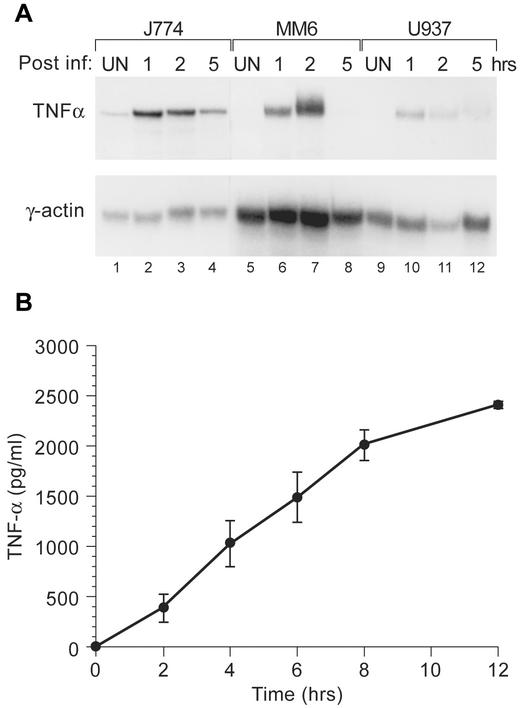
Tumor necrosis factor alpha (TNF-alpha) plays an important role in host containment of infection by Mycobacterium tuberculosis, one of the leading causes of death by an infectious agent globally. Using the pathogenic M. tuberculosis strain H37Rv, we present evidence that upon stimulation of monocytic cells by M. tuberculosis a unique TNF-alpha enhanceosome is formed, and it is distinct from the TNF-alpha enhanceosome that forms in T cells stimulated by antigen engagement or virus infection. A distinct set of activators including ATF-2, c-jun, Ets, Sp1, Egr-1 and the coactivator proteins CBP/p300 are recruited to the TNF-alpha promoter after stimulation with M. tuberculosis. Furthermore, the formation of this enhanceosome is dependent on inducer-specific helical phasing relationships between transcription factor binding sites. We also show that the transcriptional activity of CBP/p300 is potentiated by mycobacterial stimulation of monocytes. The identification of TNF-alpha regulatory elements and coactivators involved in M. tuberculosis-stimulated gene expression thus provides potential selective molecular targets in the modulation of TNF-alpha gene expression in the setting of mycobacterial infection.


Figures

FIG. 1.
FIG. 1.
H37Rv induces TNF-α transcription and protein expression. (A) An RNase protection assay was performed with three monocytic cell lines (murine J774, human MonoMac6, and human U937). Cells were infected with live M. tuberculosis H37Rv (MOI, 5:1) or mock infected. Cells were collected at 1, 2, and 5 h postinfection as indicated. Total RNA was extracted and hybridized to radiolabeled antisense RNA probes for TNF-α or actin (control) as described in Materials and Methods. (B) J774 cells were stimulated with H37Rv whole sonicate (10 μg/ml) for the times indicated, and supernatants were collected and assayed for TNF-α protein levels by using a Quantikine murine TNF-α ELISA kit. Unstimulated control cells did not produce detectable levels of TNF-α protein at any time point (data not shown). The H37Rv whole sonicate contained 0.075 pg of endotoxin/μg as determined by a kinetic, turbidimetric Limulus amoebocyte lysate assay.
FIG.2.
FIG.2.
Identification of activator binding sites required for TNF-α gene regulation by M. tuberculosis. (A) J774 cells were transfected with 1 μg of a TNF-α luciferase reporter containing either 200 or 982 nt upstream of the transcription start site. Eight hours posttransfection, cells weretreated with H37Rv M. tuberculosis whole sonicate (10 μg/ml) for 16 h. All transfections included a control Renilla luciferase plasmid and were normalized to Renilla luciferase activity. Data are presented as means ± standard errors of the means (SEM) of three independent experiments. (B) The DNA sequence of the TNF-α promoter spanning nt −200 to −20 relative to the transcription start site is shown. Transcription factor binding sites are indicated, as are the locations of point mutations in the mutant constructs studied in panel C. (C) The CRE, Sp1, upstream Sp1, Egr-1, and Ets binding sites are required for M. tuberculosis induction of TNF-α. J774 cells were transfected with 1 μg of the −200 TNF-α luciferase reporter or with isogenic reporters containing the indicated mutations and treated with M. tuberculosis whole sonicate for 16 h as described above. We noted that the 3′M and −76 NFAT mutant abolished Ets binding to the adjacent −84-Ets/Elk site (reference and data not shown). Data are presented as means ± SEM of three independent experiments. (D) Formaldehyde cross-linking and chromatin immunoprecipitation of J774 cells that were unstimulated (−) or treated with M. tuberculosis whole sonicate (+). Samples of sonicated and purified chromatin were immunoprecipitated with the indicated antibodies, and DNA isolated from immunoprecipitated material was amplified by PCR with primers specific for the TNF-α promoter. An increase in the relative amount of the TNF-α promoter-specific PCR product indicates binding of the protein to the endogenous amplified TNF-α promoter. Densitometry quantification of the induction ratios for the various transcription factors were 1.9 for ATF-2, 2.0 for c-Jun, 3.8 for Ets, 1.5 for Sp1, and 1.6 for Egr. Input DNA control lanes (lanes 12 and 13) and free primer (lane 1) are shown.
FIG. 3.
FIG. 3.
CBP/p300 proteins are required for M. tuberculosis induction of TNF-α. (A) Inhibition of CBP/p300 impairs TNF-α gene induction by M. tuberculosis. J774 cells were cotransfected with 1 μg of the −200 TNF-α luciferase reporter and with 2 μg of the vectors expressing wild-type E1A 12S or mutant (Δ2-36) forms of E1A 12S as indicated. Following transfection, cells were treated with M. tuberculosis whole sonicate for 16 h. Data are presented as means ± standard errors of the means (SEM) of three independent experiments. (B) M. tuberculosis potentiates the transcriptional activity of CBP and p300. J774 cells were cotransfected with a Gal4-dependent luciferase reporter (Gal4x5-luc, 1 μg) and increasing amounts of vectors expressing full-length CBP or full-length p300 fused to the Gal4 DNA-binding domain (0.02, 0.07, 0.2, 0.7, or 2 μg) or the Gal4 DNA-binding domain alone (2 μg). Transfected cells were then treated with M. tuberculosis whole sonicate for 16 h (Mtb) or mock stimulation (UN). Data are presented as means ± SEM of three independent experiments. DBD, DNA-binding domain.
FIG. 4.
FIG. 4.
M. tuberculosis enhanceosome formation is dependent upon helical phasing. J774 macrophages were transfected with the wild-type −200 TNF-α luciferase reporter gene or constructs with an insertion of 5 or 10 nt as indicated. These insertions add one-half of a helical turn (5 nt) and a full helical turn (10 nt) and disrupt or restore the precise helical phasing of DNA-bound transcription factors. The cells were then stimulated with M. tuberculosis H37Rv whole sonicate and the increase in inducibility (n-fold) was calculated. All transfections included a control Renilla luciferase plasmid and were normalized to Renilla luciferase activity. Data are presented as means ± standard errors of the means of four independent experiments.
FIG. 5.
FIG. 5.
A model of the recruitment of inducer-specific TNF-α enhanceosomes in T lymphocytes and monocytic cells. Distinct enhanceosomes are formed on the TNF-α promoter region in T cells in response to ionophore (top panel) or virus stimulation (middle panel) and in monocytes in response to M. tuberculosis and LPS stimulation (bottom panel). The M. tuberculosis-LPS model shows simultaneous binding of Egr-1 and of Sp1 at the upstream Sp1 site; since their binding sites have some overlap. The phasing mutants (data not shown) and site-directed mutagenesis experiments previously reported (23) gave similar results when using LPS as the stimulus, indicating that the same enhanceosome (or a similar one) is assembled after LPS as that assembled after M. tuberculosis stimulation of monocytic cells.

References

    1. Bean, A. G., D. R. Roach, H. Briscoe, M. P. France, H. Korner, J. D. Sedgwick, and W. J. Britton. 1999. Structural deficiencies in granuloma formation in TNF gene-targeted mice underlie the heightened susceptibility to aerosol Mycobacterium tuberculosis infection, which is not compensated for by lymphotoxin. J. Immunol. 162:3504-3511. - PubMed
    1. Bermudez, L. E., and L. S. Young. 1988. Tumor necrosis factor, alone or in combination with IL-2, but not IFN-gamma, is associated with macrophage killing of Mycobacterium avium complex. J. Immunol. 140:3006-3013. - PubMed
    1. Carey, M. 1998. The enhanceosome and transcriptional synergy. Cell 92:5-8. - PubMed
    1. Chrivia, J. C., R. P. Kwok, N. Lamb, M. Hagiwara, M. R. Montminy, and R. H. Goodman. 1993. Phosphorylated CREB binds specifically to the nuclear protein CBP. Nature 365:855-859. - PubMed
    1. Eckner, R., M. E. Ewen, D. Newsome, M. Gerdes, J. A. DeCaprio, J. B. Lawrence, and D. M. Livingston. 1994. Molecular cloning and functional analysis of the adenovirus E1A-associated 300-kD protein (p300) reveals a protein with properties of a transcriptional adaptor. Genes Dev. 8:869-884. - PubMed

Publication types

MeSH terms

Substances

LinkOut - more resources