. 2003 Jan;23(2):645-54.
doi: 10.1128/MCB.23.2.645-654.2003.

Elevated phospholipase D activity in H-Ras- but not K-Ras-transformed cells by the synergistic action of RalA and ARF6

Affiliations

Elevated phospholipase D activity in H-Ras- but not K-Ras-transformed cells by the synergistic action of RalA and ARF6

Lizhong Xu et al. Mol Cell Biol. 2003 Jan.

Abstract

Phospholipase D (PLD) activity is elevated in response to the oncogenic stimulus of H-Ras but not K-Ras. H-Ras and K-Ras have been reported to localize to different membrane microdomains, with H-Ras localizing to caveolin-enriched light membrane fractions. We reported previously that PLD activity elevated in response to mitogenic stimulation is restricted to the caveolin-enriched light membrane fractions. PLD activity in H-Ras-transformed cells is dependent upon RalA, and consistent with a lack of elevated PLD activity in K-Ras-transformed cells, RalA was not activated in K-Ras-transformed cells. Although H-Ras-induced PLD activity is dependent upon RalA, an activated mutant of RalA is not sufficient to elevate PLD activity. We reported previously that RalA interacts with PLD activating ADP ribosylation factor (ARF) proteins. In cells transformed by H-Ras, we found increased coprecipitation of ARF6 with RalA. Moreover, ARF6 colocalized with RalA in light membrane fractions. Interestingly, ARF6 protein levels were elevated in H-Ras- but not K-Ras-transformed cells. A dominant-negative mutant of ARF6 inhibited PLD activity in H-Ras-transformed NIH 3T3 cells. Activated mutants of either ARF6 or RalA were not sufficient to elevate PLD activity in NIH 3T3 cells; however, expression of both activated RalA and activated ARF6 in NIH 3T3 cells led to increased PLD activity. These data suggest a model whereby H-Ras stimulates the activation of both RalA and ARF6, which together lead to the elevation of PLD activity.


Figures

FIG. 1.
FIG. 1.
PLD activity is elevated in H-Ras- but not K-Ras-transformed cells. (A) Untransformed NIH 3T3 cells and NIH 3T3 cells transformed by H-Ras and K-Ras were placed in medium containing 0.5% serum for 1 day. PLD activity was then determined by the transphosphatidylation reaction in the presence of 0.8% butanol as described in Materials and Methods. The PLD activity was normalized to the PLD activity in the parental NIH 3T3 cells, which was given a value of 1. Error bars represent the standard deviation for three independent experiments performed in duplicate. (B) Cell lysates from the parental and H-Ras- and K-Ras-transformed NIH 3T3 cells were subjected to Western blot analysis with antibodies specific for H-Ras, K-Ras, and caveolin 1 as indicated. (C) NIH 3T3 cells were transiently transfected with vectors that express H-Ras, K-Ras, and the parental pZIP-NeoSV(X)1 empty vector. Twenty-four hours after transfection, the cells were placed in fresh medium containing 0.5% serum, and after an additional 24 h, PLD activity was determined as in panel A and normalized to the PLD activity in the NIH 3T3 cells that were transfected with the empty vector pZIP-NeoSV(X)1, which was given a value of 1. Ras protein levels were determined by Western blot analysis as shown.
FIG. 2.
FIG. 2.
RalA activation in H-Ras- and K-Ras-transformed cells. Untransformed NIH 3T3 cells and NIH 3T3 cellS transformed by either H-Ras or K-Ras were lysed and then treated with immobilized GST-Ral-BD as described in Materials and Methods. GST-Ral-BD was recovered by centrifugation, and the precipitate was subjected to Western blot analysis with an antibody raised against RalA. Total RalA protein levels in the cell lysates were also examined by Western blot analysis of the untreated lysates as shown. The figure shown is representative of four independent experiments. The relative level of activated RalA was determined by densitometric analysis of the Ral-BD precipitates for four independent experiments normalized to the control NIH 3T3 cells and given a value of 1. Error bars represent the standard error for the four experiments.
FIG. 3.
FIG. 3.
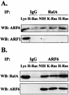
Increased coprecipitation of ARF6, but not ARF1, with RalA in H-Ras-transformed cells. (A) RalA was immunoprecipitated (IP) from lysates of parental and H-Ras- and K-Ras-transformed NIH 3T3 cells by using a mouse monoclonal RalA antibody. The RalA immunoprecipitates (RalA) were then subjected to Western blot (WB) analysis with antibodies raised against either ARF1 or ARF6 (rabbit IgGs). A nonimmune IgG control (IgG) is shown, as is a portion of whole-cell lysate (4%) (Lys) that was not subjected to immunoprecipitation. (B) A reciprocal experiment is shown in which lysates of parental and H-Ras- and K-Ras-transformed NIH 3T3 cells were immunoprecipitated with anti-ARF6 (rabbit IgG) in antigen excess and subjected to Western blot analysis with an antibody to RalA (mouse IgG). The membrane was stripped and reprobed with anti-ARF6 antibody to check the levels of ARF6 protein immunoprecipitated. The panels shown are representative of at least three independent experiments.
FIG. 4.
FIG. 4.
RalA colocalizes with ARF6 but not ARF1. Parental and H-Ras-transformed NIH 3T3 cells were disrupted by Dounce homogenization and then sonication as described in Materials and Methods. The membrane fragments were then run over a discontinuous gradient of 5, 35, and 45% sucrose. Twelve fractions and a pellet were recovered and subjected to Western blot analysis with antibodies to RalA, ARF1, ARF6, and total ARF (monoclonal antibody 1D9). The amount of material loaded onto the gels was normalized by volume from each of the fractions. The panels shown are representative of three independent experiments.
FIG. 5.
FIG. 5.
ARF6 is elevated in H-Ras- but not K-Ras-transformed cells. Homogenates from untransformed NIH 3T3 cells and NIH 3T3 cells transformed by either H-Ras, K-Ras, v-Src, or v-Raf were prepared as described in Materials and Methods and subjected to Western blot analysis with antibodies for ARF1 and ARF6. The data shown are representative of at least four independent experiments. The data from several (n) experiments were quantified by densitometry of Western blots. The level of ARF6 protein relative to that in the parental NIH 3T3 cells (Relative ARF6) was then determined with standard deviations (S.D.) as shown.
FIG. 6.
FIG. 6.
Activated ARF6 elevates PLD activity in NIH 3T3 cells expressing activated RalA. (A) NIH 3T3 cells and NIH 3T3 cells overexpressing an activated RalA mutant (Q72L) (35) were transiently transfected with vectors that express activated mutants of ARF1 (Q71L) and ARF6 (Q67L), as well as the parental pcDNA3 empty vector. Twenty-four hours after transfection, the cells were placed in fresh medium containing 0.5% serum, and after an additional 24 h, PLD activity was determined as in Fig. 1A and normalized to the PLD activity in the NIH 3T3 cells that were transfected with the empty vector, which was given a value of 1. The levels of RalA, ARF1, and ARF6 were determined by Western blot analysis as shown. (B) NIH 3T3 cells were transiently transfected with either pZIP-NeoSV(X)1 empty vector or activated RalA (Q72L) mutant, along with pcDNA3 empty vector, or with vectors expressing either the activated mutant of ARF1 (Q71L) or ARF6 (Q67L) as indicated. PLD activity was determined as in panel A and normalized to the PLD activity in the NIH 3T3 cells transfected with the empty vector controls. RalA and ARF protein levels were determined as in panel A. Error bars for panels A and B represent the standard deviation for three independent experiments performed in duplicate.
FIG. 7.
FIG. 7.
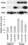
H-Ras-induced PLD activity is dependent on ARF6. Parental and H-Ras-transformed NIH 3T3 cells were transiently transfected with vectors that express dominant-negative mutants for ARF1 (T31N) and ARF6 (T27N) as described in Materials and Methods. As a control, these cells were transfected with pcDNA3 empty vector. Forty-eight hours after transfection, PLD activity was determined as in Fig. 6. The PLD activity was normalized to the PLD activity in the empty vector control for the NIH 3T3 cells, which was given a value of 1. Ras and ARF protein levels were determined as in Fig. 6. Error bars represent the standard deviation for three independent experiments performed in duplicate.
FIG. 8.
FIG. 8.
ARF6 mediates EGF-induced PLD activity. (A) EGFR cells were stably transfected with pcDNA3 (empty vector) and the vector for expression of the dominant-negative mutant ARF6 (T27N). Cells were selected in the presence of G418 for 2 weeks, and clones were pooled and used for experiments. EGF (100 ng/ml) was added as indicated, and the PLD activity was determined after 15 min. The PLD activity was normalized to the PLD activity in the empty vector control cells without EGF, which was given a value of 1. Error bars represent the standard deviation for three independent experiments performed in duplicate. The level of ARF6 proteins in the transfected cells was confirmed by Western blot (WB) analysis (B). (C) Lysates from either untreated EGFR cells or EGFR cells treated with EGF (100 ng/ml) for 15 min were lysed and then immunoprecipitated (IP) with an anti-RalA antibody. Immunoprecipitates were then subjected to Western blot analysis with antibodies to either ARF1 or ARF6 as in Fig. 3. A reciprocal experiment is shown in which cell lysates were immunoprecipitated with anti-ARF6 antibody and subjected to Western blot analysis with an antibody to RalA. The membrane was stripped and reprobed with anti-ARF6. The figure shown is a representative of at least three independent experiments.
FIG. 9.
FIG. 9.
Working model for activation of PLD1 by the synergistic action of RalA and ARF 6 in H-Ras-transformed cells. It is proposed that H-Ras activates parallel pathways leading to the activation of Ral-GDS and an as-yet-unspecified ARF-GDS. Activation of Ral-GDS activates RalA, which is already in a complex with PLD1. The activation of RalA is proposed to recruit activated ARF6 into the RalA-PLD1 complex, or alternatively activated ARF6 recruits the activated RalA-PLD1 into a RalA-PLD1-RalA complex. The activated ARF6 then activates PLD1.

Similar articles

Cited by

References

    1. Aguirre Ghiso, J., P. Frankel, Z. Lu, H. Jiang, E. Farias, A. Olsen, L. A. Feig, E. B. de Kier Joffe, and D. A. Foster. 1999. RalA requirement for v-Src- and v-Ras-induced tumorigenicity and overproduction of urokinase-type plasminogen activator and metalloproteases. Oncogene 18:4718-4725. - PubMed
    1. Alam, M. S., Y. Banno, S. Nakashima, and Y. Nozawa. 1995. Defective phospholipase D activation in Ki-ras-transformed NIH3T3 cells: evidence for downstream effector of PLC-γ1 in PDGF-mediated signal transduction. Biochem. Biophys. Res. Commun. 207:460-466. - PubMed
    1. Apolloni, A., I. A. Prior, M. Lindsay, R. G. Parton, and J. F. Hancock. 2000. H-Ras but not K-Ras traffics to the plasma membrane through the exocytic pathway. Mol. Cell. Biol. 20:2475-2487. - PMC - PubMed
    1. Bos, J. L. 1989. Ras oncogenes in human cancer: a review. Cancer Res. 49:4682-4689. - PubMed
    1. Boshans, R. L., S. Szanto, L. van Aelst, and C. D'Souza-Schorey. 2000. ADP-ribosylation factor 6 regulates actin cytoskeleton remodeling in coordination with Rac1 and RhoA. Mol. Cell. Biol. 20:3685-3694. - PMC - PubMed

Publication types