. 2003 Jan;23(2):721-32.
doi: 10.1128/MCB.23.2.721-732.2003.

Regulation of RelA (p65) function by the large subunit of replication factor C

Affiliations

Regulation of RelA (p65) function by the large subunit of replication factor C

Lisa A Anderson et al. Mol Cell Biol. 2003 Jan.

Abstract

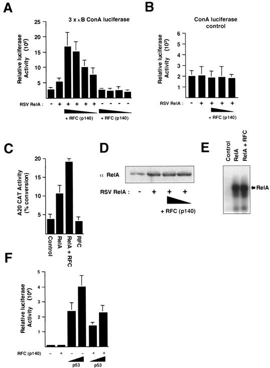
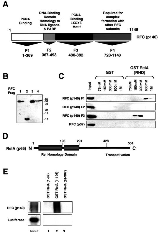
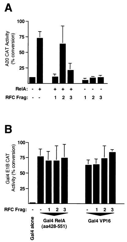
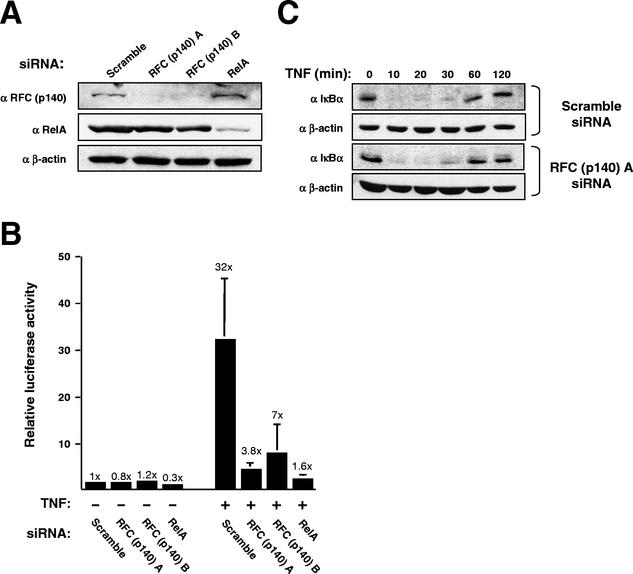
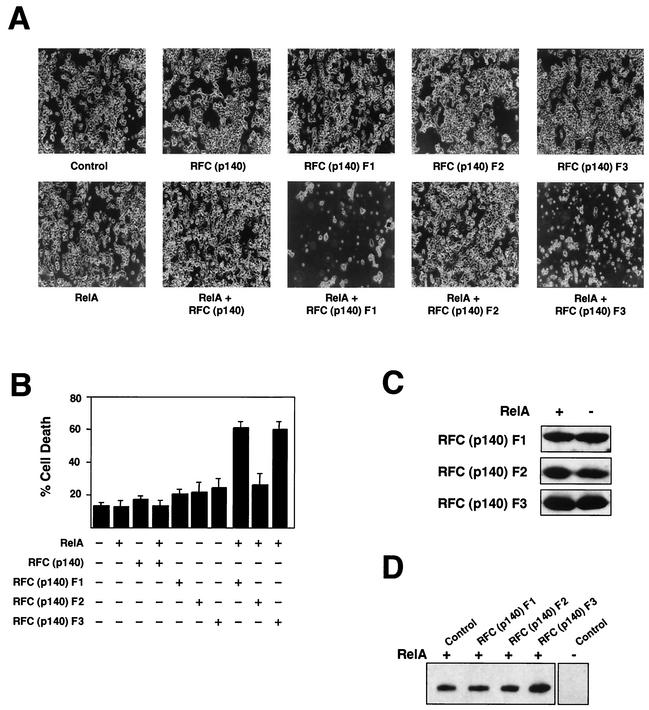
The RelA (p65) subunit of NF-kappaB is an important regulator of inflammation, proliferation, and apoptosis. We have discovered that the large subunit, p140, of replication factor C (RFC) can function as a regulator of RelA. RFC is a clamp loader, facilitating the addition and removal of proliferating-cell nuclear antigen from DNA during replication and repair but can also interact directly with the retinoblastoma tumor suppressor protein and the transcription factor C/EBPalpha. We find that RFC (p140) interacts with RelA both in vitro and in vivo and stimulates RelA transactivation. In contrast, coexpression of fragments of RFC (p140) that mediate the interaction with RelA results in transcriptional inhibition. The significance of this regulation was confirmed by using short interfering RNA oligonucleotides targeted to RFC (p140). Down regulation of endogenous RFC (p140) inhibits expression from a chromosomally integrated reporter plasmid induced by endogenous, TNF-alpha-activated NF-kappaB. Dominant negative fragments of RFC (p140) also cooperate with overexpressed RelA to induce cell death. Interestingly, RFC (p140) also interacts with the tumor suppressor p53. Taken together, these observations suggest that, in addition to its previously described function in DNA replication and repair, RFC (p140) has an important role as a regulator of transcription and NF-kappaB activity.


Figures

FIG. 1.
FIG. 1.
RFC (p140) regulates RelA transcriptional activity. (A) RFC (p140) stimulates RelA transactivation. U-2 OS cells were transfected with the 3 × κB ConA luciferase reporter plasmid (1.5 μg) and the indicated RSV RelA (1 μg) or pCDNA3 RFC (p140) expression plasmid (0.1, 0.5, 1, and 2 μg). Control RSV or pCDNA3 plasmids were included in all transfections such that each condition had the same level of each type of plasmid. Cells were harvested after 30 h. Results shown are the means of three separate experiments. Standard deviations are shown. (B) RelA and RFC (p140) do not affect the activity of a control reporter plasmid. U-2 OS cells were transfected as described for panel A, except that a control ConA luciferase reporter plasmid lacking κB elements was used. (C) RFC (p140) stimulates RelA transactivation of the A20 promoter. U-2 OS cells were transfected as described for panel A with A20 CAT reporter plasmid (5 μg) and the indicated RSV RelA (5 μg) or pCDNA3 RFC (p140) expression plasmid (0.5 μg). Results shown are the means of three separate experiments. Standard deviations are shown. (D) RFC (p140) does not affect transfected RelA protein levels. U-2 OS cells were transfected as described for panel A; however, after 30 h, whole-cell lysates were prepared and analyzed by Western blot analysis for RelA protein levels. (E) RFC (p140) does not affect RelA DNA binding. HEK 293 cells were transfected with a RelA or RFC (p140) expression plasmid (5 μg of each), either alone or in combination, as indicated. Nuclear protein extracts were prepared and analyzed by EMSA with a 32P-labeled oligonucleotide containing the Ig/HIV NF-κB binding site. The position of the RelA-DNA complex is indicated. (F) RFC (p140) represses p53 transactivation. U-2 OS cells were transfected as described for panel A with the Bax luciferase reporter plasmid (1.5 μg) and the indicated pCDNA3 p53 (100 and 500 ng) or pCDNA3 RFC (p140) (0.5 μg) expression plasmid. Results shown are the means of three separate experiments. Standard deviations are shown.
FIG. 2.
FIG. 2.
RelA and p53 interact with RFC (p140). (A) Immunoprecipitated RelA binds in vitro-translated RFC (p140). RelA was immunoprecipitated from nuclear protein extracts (200 μg) prepared from HEK 293 cells transfected with a RelA expression plasmid. The immunoprecipitated complex was then used in a pull-down assay with reticulocyte lysate-translated RFC (p140). A sample of input material (10%) is shown in this and subsequent panels. IP, immunoprecipitating. (B) RFC (p140) binds the RHD of RelA. Purified GST, GST RelA (RHD), or GST RelA (428 to 551), expressed in E. coli and bound to glutathione agarose, was used in a pull-down assay with reticulocyte lysate-translated RFC (p140). (C) Overexpressed RelA coimmunoprecipitates with RFC (p140). Endogenous RFC (p140) was immunoprecipitated from nuclear protein extracts (200 μg) prepared from HEK 293 cells transfected with either an RSV RelA expression plasmid or a control plasmid. The immunoprecipitated complex was then resolved by SDS-PAGE and immunoblotted with an anti-RelA antibody. PI, preimmune serum. (D) Endogenous RelAcoimmunoprecipitates with RFC (p140). Endogenous RFC (p140) was immunoprecipitated from U-2 OS cell nuclear protein extracts (300 μg) that had been stimulated with TNF to activate endogenous NF-κB. The immunoprecipitated complex was then resolved by SDS-PAGE and immunoblotted with an anti-RelA antibody. (E) Endogenous p53 from HEK 293 cells coimmunoprecipitates with RFC (p140). Endogenous RFC (p140) was immunoprecipitated from HEK 293 cell nuclear protein extracts (400 μg). The immunoprecipitated complex was then resolved by SDS-PAGE and immunoblotted with an anti-p53 antibody. (F) UV-activated p53 coimmunoprecipitates with RFC (p140). Endogenous RFC (p140) was immunoprecipitated from U-2 OS cell nuclear protein extracts (120 μg) that were either prepared 8 h after UV light stimulation (40 J/m2) or left untreated. The immunoprecipitated complex was then resolved by SDS-PAGE and immunoblotted with anti-p53, DO-1, antibody. (G) RFC (p140) is specifically retained in a GST RelA (RHD) column. HEK 293 cell nuclear protein extracts were passed over a GST RelA (RHD) or a GST control column. Each column was then washed before stepwise elution with buffer containing 75, 150, 300, 600, and 1,000 mM NaCl. Eluates were precipitated with TCA, resolved by SDS-PAGE, and analyzed by Western blotting with the antibodies indicated.
FIG. 3.
FIG. 3.
RelA interacts with two distinct domains of RFC (p140). (A) Schematic diagram showing different domains of RFC (p140). PARP, poly(ADP-ribose) polymerase. (B) Western blot analysis of RFC (p140) fragments (Frag) F1 to F4. Nuclear extracts were prepared from HEK 293 cells transfected with 5 μg of pCGN RFC (p140) fragment expression plasmid. Samples were resolved by SDS-PAGE and immunoblotted with anti HA-antibody 12CA5. The values on the right are molecular sizes in kilodaltons. (C) RelA interacts with RFC (p140) fragments F1 and F3. HEK 293 cell nuclear protein extracts were prepared from cells transfected with the indicated HA-tagged RFC (p140) fragments or RFC (p37). Protein affinity chromatography was then performed with the GST RelA (RHD) or control GST protein essentially as described for Fig. 2G, with 700 μg of the nuclear protein extracts. Eluates were precipitated with TCA, resolved by SDS-PAGE, and analyzed by Western blotting with an anti-HA antibody. (D) Schematic diagram showing different domains of RelA. (E) RFC (p140) binds the amino-terminal subdomain of the RelA RHD. Purified GST RelA (1 to 97), GST RelA (1 to 196), or GST RelA (61 to 307), expressed in E. coli and bound to glutathione agarose, was used in a pull-down assay with reticulocyte lysate-translated RFC (p140) or a luciferase control protein, as indicated.
FIG. 4.
FIG. 4.
Fragments (Frag) of RFC (p140) specifically repress RelA-dependent transactivation. (A) U-2 OS cells were transfected with A20 CAT reporter plasmid (5 μg) and the indicated RSV RelA (5 μg) and pCGN RFC (p140) fragment (5 μg) expression plasmids. Control RSV or cytomegalovirus plasmids were included in all transfections such that each condition had the same level of each type of plasmid. Cells were harvested after 30 h and assayed for CAT activity. Results shown are the means of three separate experiments. Standard deviations are shown. (B) Dishes (10-cm diameter) of U-2 OS cells were transfected with Gal4 E1B CAT reporter plasmid (5 μg), pCGN RFC (p140) fragment expression plasmids (5 μg), and 0.15 ng of pCDNA3 Gal4, Gal4 RelA (aa 428 to 551), and Gal4 VP16, as indicated. Control pCDNA3 plasmids were included in all transfections such that each condition had the same level of each type of plasmid. Cells were harvested after 30 h and assayed for CAT activity. Results shown are the means of three separate experiments. Standard deviations are shown.
FIG. 5.
FIG. 5.
siRNA-mediated down regulation of RFC (p140) levels inhibits endogenous NF-κB transactivation. (A) siRNAs directed against RFC (p140) and RelA specifically inhibit the expression of their target proteins. A Western blot shows the effect on the RFC (p140), RelA, and β-actin proteins of treating HeLa 57A cells with the indicated siRNAs. (B) Effect of siRNA treatment on stimulation of the integrated 3 × κB ConA luciferase reporter plasmid by TNF-α. HeLa 57A cells were treated with the indicated siRNAs. Cells were either left unstimulated or subjected to TNF-α stimulation (10 ng/ml) for 6 h, as indicated. Luciferase activity is expressed as fold activation relative to the level of activity seen in unstimulated cells with the Scramble siRNA control. The results shown are means of three separate experiments and the standard deviations together with the fold activations. (C) Treatment with anti-RFC (p140) siRNA does not affect IκBα degradation and resynthesis. HeLa 57A cells were treated as described for panel B, and whole-cell lysates were prepared and subjected to Western blot analysis with the indicated antibodies.
FIG. 6.
FIG. 6.
Disruption of RFC (p140) function results in RelA-dependent induction of cell death. (A) HEK 293 cells were transfected with the indicated pVR1012 RelA (5 μg) and pCGN RFC (p140) (5 μg) expression plasmids. Control pVR1012 or cytomegalovirus plasmids were included in all transfections such that each condition had the same level of each type of plasmid. Representative fields of view are shown. Cell death occurred between 48 and 72 h after transfection. (B) HEK 293 cells were transfected as described above with 5 μg of expression plasmids. At 72 h after transfection, cells were harvested and stained with trypan blue. Living and dead cells were then counted in triplicate with a hemocytometer. The results of two separate experiments are shown. (C and D) HEK 293 cells were transfected as described above with 5 μg of expression plasmids, and whole-cell lysates were prepared after 48 h but prior to the occurrence of cell death. Extracts were resolved by SDS-PAGE and immunoblotted with either anti-HA antibody to detect RFC protein fragment expression levels (C) or anti-RelA antibody (D).

Similar articles

Cited by

References

    1. Agalioti, T., S. Lomvardas, B. Parekh, J. M. Yie, T. Maniatis, and D. Thanos. 2000. Ordered recruitment of chromatin modifying and general transcription factors to the IFN-β promoter. Cell 103:667-678. - PubMed
    1. Anderson, L. A., and N. D. Perkins. 2002. The large subunit of replication factor C interacts with the histone deacetylase, HDAC1. J. Biol. Chem. 277:29550-29554. - PubMed
    1. Barkett, M., and T. D. Gilmore. 1999. Control of apoptosis by Rel/NF-κB transcription factors. Oncogene 18:6910-6924. - PubMed
    1. Barnes, P. J., and M. Karin. 1997. Mechanisms of disease: nuclear factor κB, a pivotal transcription factor in chronic inflammatory diseases. N. Engl. J. Med. 336:1066-1071. - PubMed
    1. Beg, A. A., W. C. Sha, R. T. Bronson, S. Ghosh, and D. Baltimore. 1995. Embryonic lethality and liver degeneration in mice lacking the RelA component of NF-κB. Nature 376:167-170. - PubMed

Publication types

MeSH terms

LinkOut - more resources