. 2003 Jan;23(2):744-53.
doi: 10.1128/MCB.23.2.744-753.2003.

Role of ALDP (ABCD1) and mitochondria in X-linked adrenoleukodystrophy

Affiliations

Role of ALDP (ABCD1) and mitochondria in X-linked adrenoleukodystrophy

M C McGuinness et al. Mol Cell Biol. 2003 Jan.

Abstract

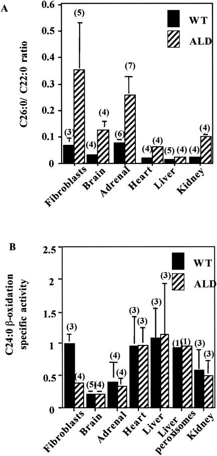
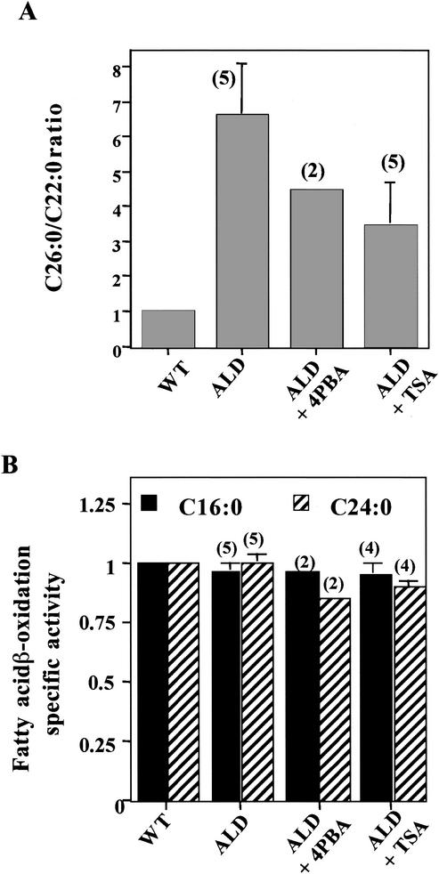
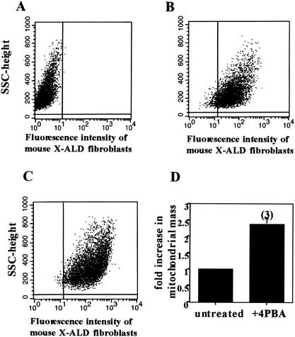
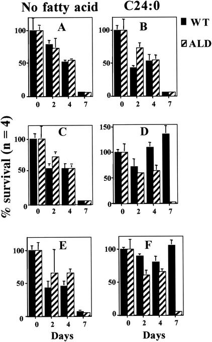
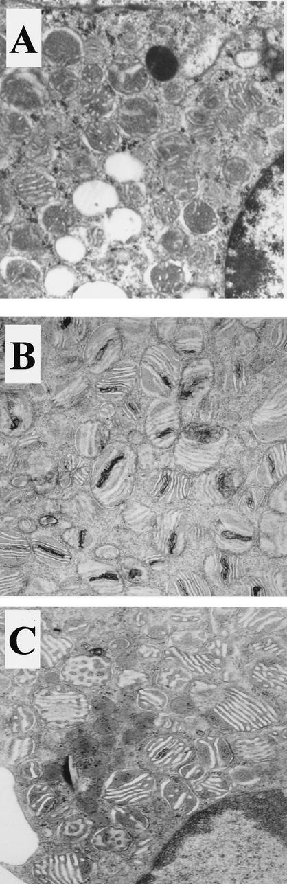
Peroxisomal disorders have been associated with malfunction of peroxisomal metabolic pathways, but the pathogenesis of these disorders is largely unknown. X-linked adrenoleukodystrophy (X-ALD) is associated with elevated levels of very-long-chain fatty acids (VLCFA; C(>22:0)) that have been attributed to reduced peroxisomal VLCFA beta-oxidation activity. Previously, our laboratory and others have reported elevated VLCFA levels and reduced peroxisomal VLCFA beta-oxidation in human and mouse X-ALD fibroblasts. In this study, we found normal levels of peroxisomal VLCFA beta-oxidation in tissues from ALD mice with elevated VLCFA levels. Treatment of ALD mice with pharmacological agents resulted in decreased VLCFA levels without a change in VLCFA beta-oxidation activity. These data indicate that ALDP does not determine the rate of VLCFA beta-oxidation and that VLCFA levels are not determined by the rate of VLCFA beta-oxidation. The rate of peroxisomal VLCFA beta-oxidation in human and mouse fibroblasts in vitro is affected by the rate of mitochondrial long-chain fatty acid beta-oxidation. We hypothesize that ALDP facilitates the interaction between peroxisomes and mitochondria, resulting, when ALDP is deficient in X-ALD, in increased VLCFA accumulation despite normal peroxisomal VLCFA beta-oxidation in ALD mouse tissues. In support of this hypothesis, mitochondrial structural abnormalities were observed in adrenal cortical cells of ALD mice.


Figures

FIG. 1.
FIG. 1.
ALD mouse tissues have normal VLCFA (C24:0) β-oxidation but accumulate VLCFA. (A) Total lipids were extracted, fractionated, and analyzed by gas chromatography as previously described (20). C26:0/C22:0 ratios are shown. These data were taken from Table 2 of reference with permission. (B) VLCFA (C24:0) β-oxidation activity (nanomoles per hour per milligram) was measured in wild-type (WT) and ALD mouse fibroblasts and tissues. Isolated peroxisomes were determined to be intact if most of the peroxisomal catalase activity was recovered in the peroxisomal pellet. Results are shown as means ± the standard deviations with the number of experiments performed in parentheses.
FIG. 2.
FIG. 2.
LCFA (C16:0) and VLCFA (C24:0) β-oxidation activities were measured in fibroblasts with a reduced capacity for mitochondrial fatty acid β-oxidation. Fibroblasts lacking either (i) CPT1, i.e., the ability to transport LCFA across the mitochondrial membrane, or (ii) (V)LCAD, i.e., the ability to metabolize LCFA, had reduced rates of LCFA (C16:0) and peroxisomal VLCFA (C24:0) β-oxidation. Fibroblasts with mutations affecting MCAD and SCAD β-oxidation had normal rates of both LCFA (C16:0) and peroxisomal VLCFA (C24:0) β-oxidation. Wild-type fatty acid β-oxidation was determined in each experiment as described in Materials and Methods, and this value was used to calculate the percentage of wild-type activity in the fibroblast cell lines being studied. Results are shown as means ± the standard deviations with the number of experiments performed in parentheses.
FIG. 3.
FIG. 3.
Effect of LCFA concentration on VLCFA (C24:0) β-oxidation and phytanic acid α-oxidation in wild-type (WT) and ALD mouse liver postnuclear supernatants (PNS) and VLCFA (C24:0) β-oxidation in isolated peroxisomes. Increasing amounts of unlabeled LCFA (C16:0) were added to mouse liver postnuclear supernatants and isolated peroxisomes, and peroxisomal VLCFA (C24:0) β-oxidation and phytanic acid α-oxidation were measured.
FIG. 4.
FIG. 4.
Effect of the mitochondrial complex III inhibitor antimycin A on fibroblast fatty acid β-oxidation. Human and mouse fibroblasts from wild-type (WT) and affected individuals were grown for 2 days in medium containing antimycin A (0.4 μg/ml). Wild-type LCFA β-oxidation (C16:0) and VLCFA (C24:0) fatty acid β-oxidation were determined in each experiment as described in Materials and Methods and set to 1. These values were used to calculate the fold decrease in activity in the fibroblast cell lines being studied. Results are shown as means ± the standard deviations with the number of experiments performed in parentheses.
FIG. 5.
FIG. 5.
Effect of 4PBA on fatty acid metabolism in vivo. VLCFA accumulation (A) and β-oxidation (B) were determined in liver postnuclear supernatants from wild-type (WT) mice, ALD mice, and ALD mice treated with 4PBA (0.16 g/kg/day) or TSA (0.3 mg/kg/day) in water. Total lipids were extracted, fractionated, and analyzed by gas chromatography as previously described (20). Wild-type mouse C26:0/C22:0 ratios and fatty acid β-oxidation activity levels were set to 1, and these values were used to calculate changes in ratios and activity levels in untreated and treated ALD mice. Results are shown as means ± the standard deviations with the number of experiments performed in parentheses.
FIG. 6.
FIG. 6.
Time course of response to 4PBA. Fibroblasts from ALD mice were grown in medium containing 5 mM 4PBA for the times indicated. LCFA (C16:0) and VLCFA (C24:0) β-oxidation activities were measured, and values were expressed as fold increases relative to untreated-control values. 4PBA treatment (5 mM) increased LCFA (C16:0) before VLCFA (C24:0) β-oxidation in ALD mouse fibroblasts.
FIG. 7.
FIG. 7.
Effect of 4PBA treatment (5 mM) on VLCFA (C24:0) β-oxidation specific activity in mitochondrial fatty acid metabolism mutants. Fibroblasts with mutations affecting CPT1, (V)LCAD, MCAD, and SCAD were exposed to 4PBA for 3 days. Cells lacking the ability to either transport LCFA across the mitochondrial membrane (CPT1) or metabolize LCFA [(V)LCAD] did not respond to 4PBA. However, MCAD- and SCAD-deficient cells, with normal LCFA metabolism, responded to 4PBA with increased LCFA (C16:0) and peroxisomal VLCFA (C24:0) β-oxidation activity. Results are shown as means ± the standard deviations with the number of experiments performed in parentheses.
FIG. 8.
FIG. 8.
Effect of 4PBA (5 mM) on mitochondrial mass in human X-ALD fibroblasts. Mitotracker Green FM was used to measure mitochondrial mass in fibroblasts by flow cytometry before and after 4PBA treatment. The autofluorescence of unstained cells (A) was analyzed and subtracted from the analysis of fibroblasts stained with Mitotracker before (B) and after (C) 4PBA treatment. The 2.4-fold increase in mitochondrial mass after 4PBA treatment is shown in panel D. SSC, single scanned cell. Results are shown as means ± the standard deviations with the number of experiments performed in parentheses.
FIG. 9.
FIG. 9.
Effects of 4PBA and TSA on mouse fibroblast VLCFA (C24:0) auxotrophy. Wild-type (WT) and ALD mouse fibroblasts were grown in glucose-free, delipidated medium (minimal medium) (A, C, and E) or in minimal medium supplemented with C24:0 (1 nmol/ml of medium) (B, D, and F) either alone (A and B) or in the presence of 4PBA (5 mM) (C and D) or TSA (50 nM) (E and F). Results are shown as means ± the standard deviations with the number of experiments performed in parentheses.
FIG. 10.
FIG. 10.
Effect of in vivo 4PBA treatment on mitochondrial ultrastructure in adrenocortical cells from ALD mice. Adrenal glands were harvested from wild-type (A), ALD (B), and 4PBA-treated ALD (C) mice after perfusion with 4% glutaraldehyde. They were postfixed in osmium tetroxide and embedded in epoxy resin. The embedded tissues were thin sectioned and stained with uranyl acetate-lead citrate and examined under an electron microscope (magnification, ×48,750).

References

    1. Allen, J., T. Kepic, D. Garwicki, and M. Yunus. 1982. Adrenal defect in adrenomyelodystrophy. South. Med. J. 75:877-879. - PubMed
    1. Baumgart, E., I. Vanhorebeek, M. Grabenbauer, M. Borgers, P. E. Declercq, H. D. Fahimi, and M. Baes. 2001. Mitochondrial alterations caused by defective peroxisomal biogenesis in a mouse model for Zellweger syndrome (PEX5 knockout mouse). Am. J. Pathol. 159:1477-1494. - PMC - PubMed
    1. Berger, J., S. Albet, M. Bentejac, N. Netik, A. Holzinger, A. A. Roscher, M. Bugaut, and S. Forss-Petter. 1999. The four murine peroxisomal ABC-transporter genes differ in constitutive, inducible and developmental expression. Eur. J. Biochem. 265:719-727. - PubMed
    1. Bezman, L., A. Moser, G. Raymond, P. Rinaldo, P. Watkins, K. Smith, N. Kass, and H. Moser. 2001. Adrenoleukodystrophy: incidence, new mutation rate, and results of extended family screening. Ann. Neurol. 49:512-517. - PubMed
    1. Bissler, J. J., M. Tsoras, H. H. H. Goring, P. Hug, G. Chuck, E. Tombragel, C. McGraw, J. Schlotman, M. A. Ralston, and G. Hug. 2002. Infantile dilated X-linked cardiomyopathy, G4.5 mutations, altered lipids, and ultrastructural malformations of mitochondria in heart, liver, and skeletal muscle. Lab. Investig. 82:335-344. - PubMed

Publication types

MeSH terms

Substances

LinkOut - more resources