. 2003 Jan 1;546(Pt 1):77-87.
doi: 10.1113/jphysiol.2002.030692.

Distribution, subcellular localization and ontogeny of ASIC1 in the mammalian central nervous system

Affiliations

Distribution, subcellular localization and ontogeny of ASIC1 in the mammalian central nervous system

Diego Alvarez de la Rosa et al. J Physiol. .

Abstract

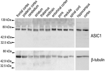
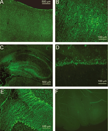
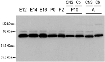
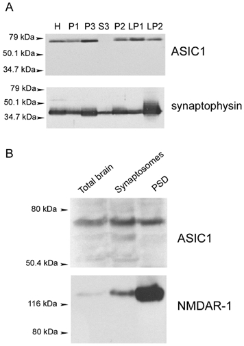
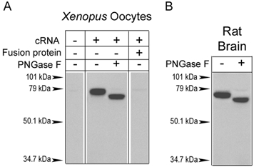
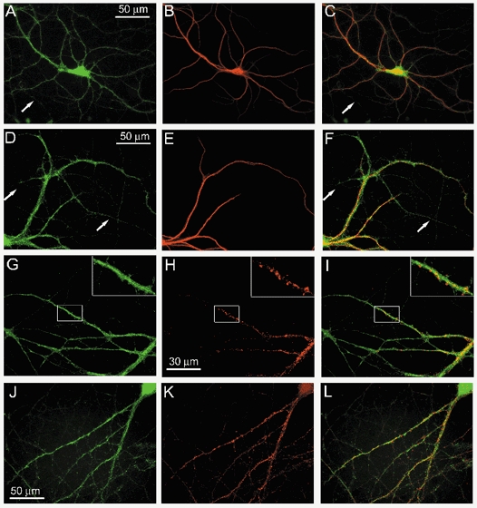
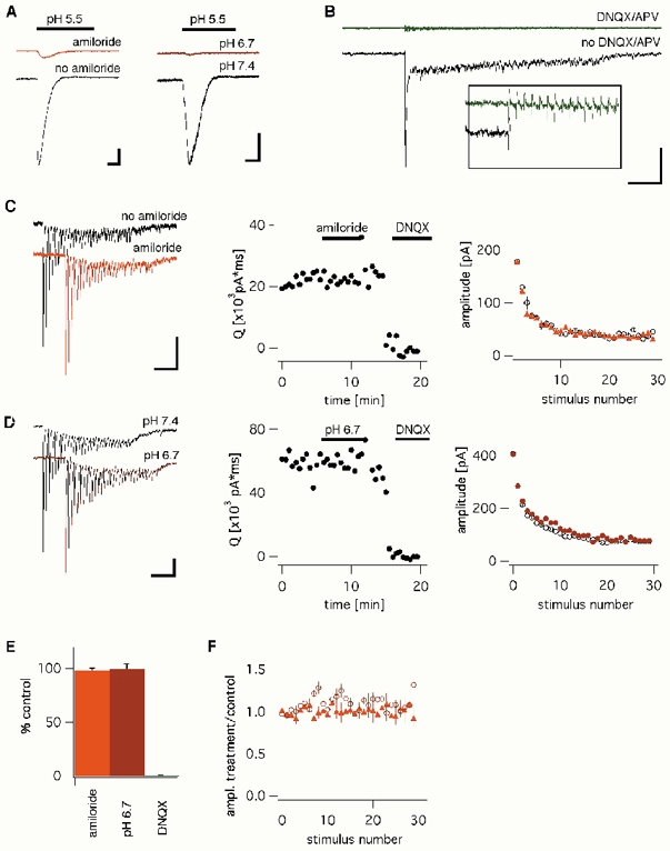
The acid-sensitive ion channel ASIC1 is a proton-gated ion channel from the mammalian nervous system. Its expression in sensory neurons and activation by low extracellular pH suggest that ASIC is involved in transmitting nociceptive impulses produced by the acidification caused by injury or inflammation. However, ASIC1 expression is not restricted to sensory neurons. To understand the functional role of ASIC1 in the CNS we investigated its expression and subcellular distribution therein. In particular, we examined the presence of ASIC1 in domains where the local pH may drop sufficiently to activate ASIC1 under physiological conditions. Immunostaining with specific antibodies revealed broad expression of ASIC1 in many areas of the adult rat brain including the cerebral cortex, hippocampus and cerebellum. Within cells, ASIC1 was found predominantly throughout the soma and along the branches of axons and dendrites. ASIC1 was not enriched in the microdomains where pH may reach low values, such as in synaptic vesicles or synaptic membranes. Pre- or postsynaptic ASIC1 was not gated by synaptic activity in cultured hippocampal neurons. Blockage or desensitization of ASIC1 with amiloride or pH 6.7, respectively, did not modify postsynaptic currents. Finally, the ontogeny of ASIC1 in mouse brain revealed constant levels of expression of ASIC1 protein from embryonic day 12 to the postnatal period, indicating an early and almost constant level of expression of ASIC1 during brain development.


Figures

Figure 1
Figure 1. Western blot analysis of acid-sensitive ion channel (ASIC)1 expression in the rat CNS
Protein extracts from the indicated areas of rat CNS (100 μg) were analysed by Western blot using consecutively anti-ASIC1-CT (upper panel) and anti-β-tubulin (lower panel) antibodies. ASIC1 immunoreactivity was present in all areas examined. Molecular weight markers are shown on the left.
Figure 2
Figure 2. Immunohistochemical staining of endogenous ASIC1 in the rat brain and spinal cord
Brain sections (10 μm thick) were immunostained with anti-ASIC1-CT antibody. A, area of brain cortex exhibiting staining of most pyramidal cells. B, same region as in A but at a higher magnification, showing labelling of axons and dendrites. C, low-magnification sagittal view of the whole hippocampus showing immunoreactivity in all hippocampal regions. D, hippocampal neurons from the CA1 region at higher magnification. E, view of the cerebellar cortex showing Purkinje cells stained over the soma and dendritic tree. F, section of cerebellum stained with anti-ASIC-CT antibody pre-incubated with an excess of the cognate fusion protein.
Figure 3
Figure 3. ASIC1 expression during mouse CNS development
Protein extracts from mouse CNS at different embryonic (E) or postnatal (P) days were collected and analysed for ASIC1 expression by Western blot. A sample from adult animals was included (A). P10 and adult samples were obtained either from cerebellum (Cb) or from total CNS. Molecular weight markers are show on the left.
Figure 4
Figure 4. Subcellular distribution of ASIC1 examined by differential fractionation of membranes from rat brain
A, synaptic vesicle preparation. Equal amounts of proteins from each fraction of synaptic vesicle preparation were loaded onto an SDS-PAGE system and analysed by Western blot with anti-ASIC1-CT and with anti-synaptophysin antibodies. H = whole homogenate, P2 = second pellet from a 9200 g spin, P3 = pellet from 184 000 g spin, S3 = supernatant from 184 000 g spin, LP1 = pellet from hypotonically lysed synaptosomes centrifuged at 25 000 g, LP2 = crude synaptic vesicle fraction. B, PSD preparation. Equal amounts of proteins from rat CNS (total brain), synaptosomes or PSD fractions were analysed by Western blot using consecutively anti-ASIC1-CT and anti-NMDA-receptor type I (NMDAR-1) antibodies.
Figure 5
Figure 5. Characterization of the anti-ASIC1-EL antibody
Western blot analysis of proteins extracted from ASIC1 cRNA-injected oocytes (A) or rat brain (B) probed with anti-ASIC1-EL antibody. Proteins in lanes 3 (A) and 2 (B) were pretreated with PNGase-F. No signal was detected in water-injected oocytes or after competition of the antibody with the cognate peptide. Single bands of ≈70 kDa were detected in ASIC1-injected oocytes and in adult rat brain. Treatment with PNGase-F reduced the molecular weight by ≈4 kDa, which is consistent with the removal of two core N-linked oligosaccharides. The migration of molecular weight markers is indicated by arrowheads.
Figure 6
Figure 6. Distribution of ASIC and colocalization with synaptic markers in cultured embryonic cortical and hippocampal neurons
A and B, co-staining of cortical neurons with anti-ASIC1-CT (A, green) and MAP2 (B, red) antibodies. C, overlay of A and B indicates that ASIC1 is expressed on the soma, dendrites and axons, whereas MAP2 is present predominantly in the dendrites and in the soma. Arrows show axons stained exclusively with ASIC1. D-F, co-staining of cortical neurons with anti-ASIC1-EL (D, green) and MAP2 (E, red), showing that both ASIC1 antibodies yield an identical pattern of staining (F, overlay). G and H, co-staining of cortical neurons with anti-ASIC1-CT (G, green) and anti-PSD-95 (H, red). I, overlay of G and H. Insets show a dendrite at high magnification showing synapses intensely labelled by PSD-95 and little overlap with ASIC1. Co-staining of hippocampal neurons with anti-ASIC-CT (J, green) and anti-PSD-95 (K, red). L, overlay of signals shown in J and K.
Figure 7
Figure 7. Effect of synaptic transmission on ASIC1 activation
A, inward currents elicited in cultured hippocampal neurons by rapid application of acidic saline (pH 5.5) in the presence or absence of 50 μm amiloride (left panel) and at a bath pH of 6.7 or 7.4 (right panel). Scale bars 0.2 nA, 2 s. B, postsynaptic current elicited by high-frequency stimulation (3 s at 100 Hz) of a presynaptic glutamatergic neuron (bottom) is completely blocked by application of 10 μm 6,7-dinitroquinoxaline-2,3-dione (DNQX) and 50 μm [d, l]-2-amino-5-phosphonovalerate (APV, average of four traces, top). The inset shows a higher-magnification view of this trace during the first 15 stimuli of the train. Scale bar: 50 pA (inset 10 pA), 1 s (inset 100 ms). C, EPSCs produced in response to a 30 Hz, 1 s stimulus train in the presence or absence of amiloride (50 μm, left panel). Integrated postsynaptic currents elicited by the stimulus trains (middle panel) and average amplitudes of EPSCs to successive stimuli within the train (right panel) are shown. D, EPSCs in response to a 30 Hz stimulus in at a bath pH of 7.4 or 6.7. E, normalized integrated postsynaptic currents during amiloride application (98.2 ± 2.0 % of pre-application currents, n = 3), at a bath pH of 6.7 (99.5 ± 4.4 %; n = 3) or after application of DNQX and APV (0.4 ± 0.3 %, n = 3). F, postsynaptic currents recorded in response to successive stimuli within a train. Current amplitudes of EPSCs after application of amiloride or at pH 6.7 have been normalized to respective current amplitudes under control conditions.

References

    1. Alvarez de la Rosa D, Zhang P, Shao D, White F, Canessa CM. Functional implications of the localization and activity of acid-sensitive channels in rat peripheral nervous system. Proc Natl Acad Sci U S A. 2002;99:2326–2331. - PMC - PubMed
    1. Andreasen M, Lambert JD, Jensen MS. Effects of new non-N-methyl-d-aspartate antagonists on synaptic transmission in the in vitro rat hippocampus. J Physiol. 1989;414:317–336. - PMC - PubMed
    1. Bahn S, Volk B, Wisden W. Kainate receptor gene expression in the developing rat brain. J Neurosci. 1994;14:5525–5547. - PMC - PubMed
    1. Baron A, Waldmann R, Lazdunski M. ASIC-like, proton-activated currents in rat hippocampal neurons. J Physiol. 2002;539:485–494. - PMC - PubMed
    1. Benson CJ, Xie J, Wemmie JA, Price MP, Henss JM, Welsh MJ, Snyder PM. Heteromultimers of DEG/ENaC subunits form H+-gated channels in mouse sensory neurons. Proc Natl Acad Sci U S A. 2002;99:2338–2343. - PMC - PubMed

Publication types

MeSH terms

LinkOut - more resources