. 2003 Jan;15(1):5-18.
doi: 10.1105/tpc.006973.

LEAFY COTYLEDON1-LIKE defines a class of regulators essential for embryo development

Affiliations

LEAFY COTYLEDON1-LIKE defines a class of regulators essential for embryo development

Raymond W Kwong et al. Plant Cell. 2003 Jan.

Abstract

Arabidopsis LEAFY COTYLEDON1 (LEC1) is a critical regulator required for normal development during the early and late phases of embryogenesis that is sufficient to induce embryonic development in vegetative cells. LEC1 encodes a HAP3 subunit of the CCAAT binding transcription factor. We show that the 10 Arabidopsis HAP3 (AHAP3) subunits can be divided into two classes based on sequence identity in their central, conserved B domain. LEC1 and its most closely related subunit, LEC1-LIKE (L1L), constitute LEC1-type AHAP3 subunits, whereas the remaining AHAP3 subunits are designated non-LEC1-type. Similar to LEC1, L1L is expressed primarily during seed development. However, suppression of L1L gene expression induced defects in embryo development that differed from those of lec1 mutants, suggesting that LEC1 and L1L play unique roles in embryogenesis. We show that L1L expressed under the control of DNA sequences flanking the LEC1 gene suppressed genetically the lec1 mutation, suggesting that the LEC1-type B domains of L1L and LEC1 are critical for their function in embryogenesis. Our results also suggest that LEC1-type HAP3 subunits arose from a common origin uniquely in plants. Thus, L1L, an essential regulator of embryo development, defines a unique class of plant HAP3 subunits.


Figures

Figure 1.
Figure 1.
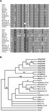
Analysis of Arabidopsis HAP3 Subunits. Amino acid sequence alignment of AHAP3 proteins. Residues highlighted in black and gray represent identical and similar amino acids, respectively. B-domain residues shared between LEC1 (At1g21970) and L1L (At5g47670) but not with the other proteins are highlighted in red. The B domain is underlined.
Figure 2.
Figure 2.
L1L RNA Is Detected Predominantly in Developing Siliques. (A) Analysis of L1L and LEC1 RNA levels with RNA gel blot hybridization experiments. Each lane contained 1 μg of poly(A) RNA from siliques with zygote- to early-globular-stage seeds (S1), siliques with globular- to heart-stage seeds (S2), siliques with torpedo- to bent-cotyledon-stage seeds (S3), siliques with mature green seeds (S4), 2-day-old seedlings (Sl), mature rosette leaves (Le), 3-week-old seedling roots (Ro), stems (St), and unopened floral buds and inflorescences (Fl). Control represents the accumulation of a ribosomal protein RNA. (B) RT-PCR amplification of L1L RNA. Abbreviations are as in (A) with the following additions: ND, no DNA; GD, wild-type genomic DNA; and lec1-1, mutant siliques with torpedo- to bent-cotyledon-stage embryos.
Figure 3.
Figure 3.
In Situ Detection of L1L RNA in Developing Embryos. Wild-type seed sections were hybridized with an L1L-specific antisense probe. All sections were exposed for 10 days. (A) to (D) and (I) to (L) show bright-field micrographs, and (E) to (H) and (M) to (P) show dark-field micrographs. The sense RNA control did not bind appreciably with the sections. Bars = 50 μm. (A) and (E) Globular-stage embryo. (B) and (F) Heart-stage embryo. (C) and (G) Linear cotyledon-stage embryo. (D) and (H) Early bent-cotyledon-stage embryo. (I) and (M) Bent-cotyledon-stage embryo. (J) and (N) Late bent-cotyledon-stage embryo. (K) and (O) Mature green-stage embryo. (L) and (P) Mature yellowing-stage embryo.
Figure 4.
Figure 4.
RNAi Suppression of L1L Gene Expression Induces Embryo Defects. (A) Seed with a wild-type embryo at the bent-cotyledon stage. The seed was cleared and viewed with Nomarski optics. (B) to (D) Cleared seeds containing defective embryos from lines containing the L1L RNAi constructs. Progeny segregating with a wild-type phenotype in the same silique were at the bent-cotyledon stage. (E) to (H) L1L RNA accumulation in defective embryos. Sections were hybridized with an antisense L1L probe and exposed for 10 days. (E) and (F) Bright- and dark-field micrographs of a defective embryo from a line containing the L1L RNAi construct. (G) and (H) Bright- and dark-field micrographs of a wild-type embryo at the mature green stage. Bars = 50 μm in (A), (D), (E), and (H) and 25 μm in (B) and (C).
Figure 5.
Figure 5.
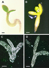
Suppression of the lec1 Mutation by L1L. (A) A representative lec1-1 seedling containing the LEC1:L1L:LEC1 transgene that has survived seed desiccation. The transgene confers desiccation tolerance to lec1 mutant embryos. (B) A lec1-1 seedling transformed with 35S:L1L. The transgene allows lec1-1 mutant embryos to withstand desiccation and confers embryonic characteristics to the seedling, including a lack of cotyledon expansion, failure of hypocotyls and roots to extend, and production of cotyledon-like organs at the positions of leaves. (C) and (D) Hybridization of cruciferin storage protein and oleosin probes, respectively, with embryo-like 35S:L1L seedlings. Sections were exposed for 2 days. Bars = 300 μm in (A) and (B) and 100 μm in (C) and (D).
Figure 6.
Figure 6.
PcL1L RNA Is Present Primarily in Developing Seeds. (A) Gel blot analysis of PcL1L RNA accumulation. Twenty-five micrograms of total RNA was analyzed from leaves (Le), stems (St), 2-week-old seedling leaves (Sl), 2-week-old seedling roots (SlRo), 2-week-old seedling stems (SlSt), inflorescences (In), ovules (Ov), 2-DAP seeds (Se I), 4- to 5-DAP seeds (Se II), 6-DAP seeds (Se III), 12- to 14-DAP embryos (Em I), and 19- to 21-DAP embryos (Em II). (B) RT-PCR analysis of PcL1L RNA accumulation. Each lane corresponds to the RNA gel blot sample in (A). (C) to (F) Distribution of PcL1L RNA. Sections were hybridized with a PcL1L antisense probe (C) to (E) or a sense RNA control (F). (C) and (D) were exposed for 4 days, whereas (E) was exposed for 47 days. (C) Preglobular-stage seed. PcL1L RNA is high in the embryo proper and suspensor. (D) Globular-stage seed. PcL1L RNA is at its highest levels in outer tissue layers of the embryo. (E) Unfertilized ovule. PcL1L RNA is present at low levels throughout the ovule. (F) Unfertilized ovule that does not bind sense RNA probe. Bars = 100 μm.
Figure 7.
Figure 7.
Identification of L1L Proteins from Other Plants. (A) Amino acid sequence alignment of the B domains of plant L1L proteins. Conserved amino acid residues are highlighted in gray, and residues unique to L1L proteins are highlighted in black. Accession numbers are given at the end of Methods. (B) Phylogenetic relationships between L1L and non-LEC1-type- HAP3 subunits. The cladogram illustrates the most parsimonious consensus pattern of relationships obtained using maximum parsimony analysis. Bootstrap values generated with 1000 replicates are indicated before the nodes. Nodes with bootstrap scores of <50% are not shown. The high bootstrap values provide strong support for the monophyletic L1L clade.

Similar articles

Cited by

References

    1. Albani, D., and Robert, L.S. (1995). Cloning and characterization of a Brassica napus gene encoding a homologue of the B subunit of a heteromeric CCAAT-binding factor. Gene 167, 209–213. - PubMed
    1. Ausubel, F.M., Brent, R., Kingston, R.E., Moore, D.D., Seidman, J.G., and Smith, J.A. (1995). Short Protocols in Molecular Biology, 3rd ed. (New York: John Wiley & Sons).
    1. Bechtold, N., Ellis, J., and Pelletier, G. (1993). In planta Agrobacterium mediated gene transfer by infiltration of adult Arabidopsis thaliana plants. C. R. Acad. Sci. Paris 316, 1194–1199.
    1. Bewley, J.D. (1997). Seed germination and dormancy. Plant Cell 9, 1055–1066. - PMC - PubMed
    1. Coustry, F., Maity, S.N., Sinha, S., and de Crombrugghe, B. (1996). The transcriptional activity of the CCAAT-binding factor CBF is mediated by two distinct activation domains, one in the CBF-B subunit and the other in the CBF-C subunit. J. Biol. Chem. 272, 14485–14491. - PubMed

Publication types

MeSH terms

Associated data