. 2003 Jan;15(1):47-62.
doi: 10.1105/tpc.006536.

Oxidative remodeling of chromoplast carotenoids: identification of the carotenoid dioxygenase CsCCD and CsZCD genes involved in Crocus secondary metabolite biogenesis

Affiliations

Oxidative remodeling of chromoplast carotenoids: identification of the carotenoid dioxygenase CsCCD and CsZCD genes involved in Crocus secondary metabolite biogenesis

Florence Bouvier et al. Plant Cell. 2003 Jan.

Abstract

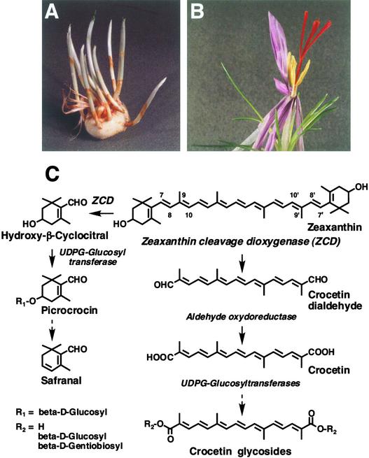
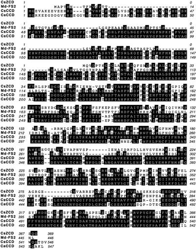
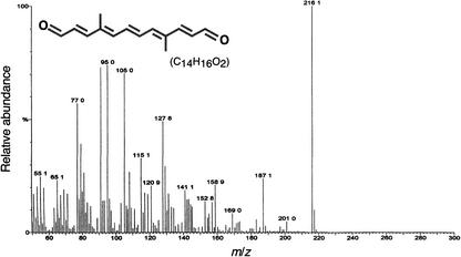
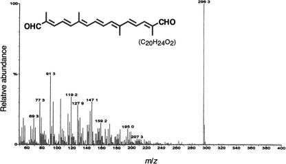
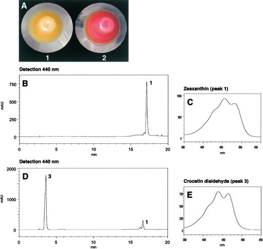
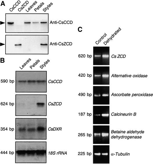
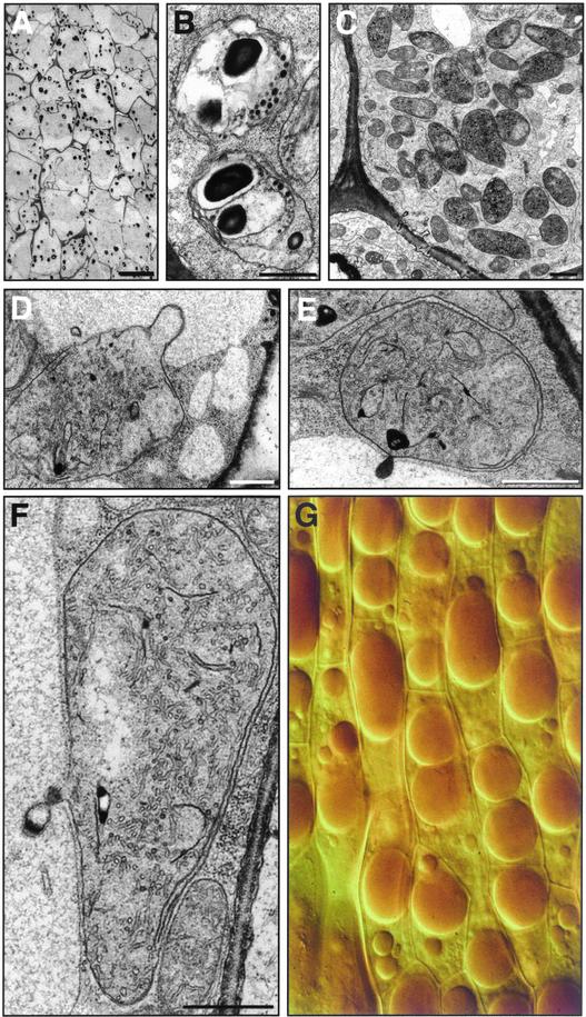
The accumulation of three major carotenoid derivatives-crocetin glycosides, picrocrocin, and safranal-is in large part responsible for the color, bitter taste, and aroma of saffron, which is obtained from the dried styles of Crocus. We have identified and functionally characterized the Crocus zeaxanthin 7,8(7',8')-cleavage dioxygenase gene (CsZCD), which codes for a chromoplast enzyme that initiates the biogenesis of these derivatives. The Crocus carotenoid 9,10(9',10')-cleavage dioxygenase gene (CsCCD) also has been cloned, and the comparison of substrate specificities between these two enzymes has shown that the CsCCD enzyme acts on a broader range of precursors. CsZCD expression is restricted to the style branch tissues and is enhanced under dehydration stress, whereas CsCCD is expressed constitutively in flower and leaf tissues irrespective of dehydration stress. Electron microscopy revealed that the accumulation of saffron metabolites is accompanied by the differentiation of amyloplasts and chromoplasts and by interactions between chromoplasts and the vacuole. Our data suggest that a stepwise sequence exists that involves the oxidative cleavage of zeaxanthin in chromoplasts followed by the sequestration of modified water-soluble derivatives into the central vacuole.


Figures

Figure 1.
Figure 1.
Accumulation and Pathway of Carotenoid-Derived Metabolites in Crocus. (A) Perianth tubes sprouting from the corm. (B) Crocus flower displaying red style branches that accumulate carotenoid-derived metabolites. (C) Possible pathway for the biosynthesis of carotenoid metabolites in Crocus style branches. Zeaxanthin, the postulated precursor, is cleaved and modified by different enzymes, which are listed in italics.
Figure 2.
Figure 2.
HPLC Analysis of Carotenoid-Derived Metabolites from Crocus Style Branches. (A) HPLC analysis of Crocus style extracts. Compounds were detected by UV-light absorbance at 220 nm. Peak 1 corresponds to picrocrocin, and peaks 2 to 4 correspond to crocetin carrying different glycosyl residues. (B) Online diode-array spectrum of picrocrocin. For peaks 2 to 4, see (D). (C) HPLC analysis of carotenoid-derived metabolites from Crocus style. Compounds were detected by visible light absorbance at 440 nm. Peaks 2 to 4 correspond to crocetin carrying different glycosyl residues. (D) Typical online diode-array spectrum of crocetin glycoside (peak 2). The profiles of peaks 3 and 4 were similar. Chromatographic separation was performed using gradient I (see Methods). mAU, milliabsorbance units.
Figure 3.
Figure 3.
Comparison of the Predicted Amino Acid Sequences of Crocus Carotenoid Cleavage Dioxygenases and Related Proteins. CsZCD and CsCCD are carotenoid cleavage dioxygenases from Crocus. Md-FS2 is an apple flower protein of unknown function (Watillon et al., 1998). CaCCD is a pepper homolog of Arabidopsis cleavage dioxygenase (Schwartz et al., 2001). Identical amino acids are indicated with black backgrounds.
Figure 4.
Figure 4.
Immunohistochemical Localization of Carotenoid Cleavage Dioxygenases in Crocus Style Branches. (A) and (B) Style sections incubated with anti-CsCCD antibody (A) and preimmune CsCCD serum (B). Bars = 50 μm. (C) and (D) Style sections incubated with anti-CsZCD antibody (C) and preimmune CsZCD serum (D). Bars = 5 μm. The images were obtained using confocal laser-scanning microscopy.
Figure 5.
Figure 5.
Functional Analysis of Recombinant CsCCD Reaction Products. (A) SDS-PAGE analysis of affinity-purified CsCCD. Soluble protein from bacteria grown at 20°C (lane 1) and 37°C (lane 2) and insoluble protein from bacteria grown at 20°C (lane 3) and 37°C (lane 4) were examined. Affinity-purified fusion protein was loaded in lane 5 (arrowhead). MW, molecular mass. (B) to (E) HPLC results (monitored at 375 nm [B] and 440 nm [D]) and online diode-array spectra of CsCCD reaction products. The separation of zeaxanthin (peak 1) and its cleavage derivative (peak 2) and online diode-array spectra of peak 2 (C) and zeaxanthin (E) are shown. mAU, milliabsorbance units. (F) to (H) UV-visible light spectra of the reaction product (peak 2) in hexane (F) and in ethanol before (G) and after (H) reduction with sodium borohydride.
Figure 6.
Figure 6.
Full-Scan Mass Spectrometry of the Reaction Product of CsCCD. Electron-impact mass spectrum of the reaction product (peak 2) obtained as shown in Figure 5B. m/z, mass-to-charge ratio.
Figure 7.
Figure 7.
Functional Analysis of Recombinant CsZCD. (A) SDS-PAGE analysis of affinity-purified CsZCD. Soluble protein from bacteria grown at 20°C without (lane 1) or after (lane 2) induction was examined. Lane 3 was loaded with affinity-purified CsZCD fusion protein (arrowhead). MW, molecular mass. (B) Thin layer chromatography analysis of CsZCD reaction products. Lane 1, incubation using soluble proteins from E. coli harboring empty vector (pBAD/TOPO ThioFusion) and zeaxanthin (Zea); lanes 2 to 4, incubations using purified recombinant CsZCD and zeaxanthin, cis-violaxanthin (cis-Viol), and trans-violaxanthin (trans-Viol). Plates were sprayed with acidic dinitrophenylhydrazine to reveal the presence of carbonyl groups. The position of the reaction product is indicated by the arrowhead. As a result of the acidic conditions, 5,6-epoxy-carotenoids were converted to 5,8-epoxy-carotenoids, which appear as green to blue spots. OR refers to the origin. (C) to (E) HPLC results and online diode-array spectra of CsZCD reaction products. The separation (monitored at 440 nm [C]) of zeaxanthin (peak 1) and its cleavage derivative (peak 3) and online diode-array spectra of peak 3 (D) and zeaxanthin (E) are shown. mAU, milliabsorbance units. (F) to (H) UV-visible light spectra of the reaction product (peak 3) in hexane (F) and in ethanol before (G) and after (H) reduction with sodium borohydride.
Figure 8.
Figure 8.
Full-Scan Mass Spectrometry of the Reaction Product of CsZCD. Electron-impact mass spectrum of the reaction product (peak 3) obtained as shown in Figure 7C. m/z, mass-to-charge ratio.
Figure 9.
Figure 9.
Plasmid-Based Assay of CsZCD in E. coli. (A) Pellet of E. coli harboring the pCAR25delB plasmid, which allows zeaxanthin production alone (1) and after coexpression of CsZCD cDNA (2). (B) to (E) HPLC results (monitored at 440 nm) and online diode-array spectra of the total lipid extract from E. coli harboring pCAR25delB plasmid ([B] and [C]) and coexpressing CsZCD cDNA ([D] and [E]). mAU, milliabsorbance units.
Figure 10.
Figure 10.
Molecular Analysis of CsCCD and CsZCD in Different Tissues of Crocus and during Dehydration Stress. (A) Protein gel blot analysis of anti-CsCCD and anti-CsZCD antibody specificity and tissue-specific expression of CsCCD and CsZCD. (B) RNA gel blot analysis and tissue-specific expression of CsCCD, CsZCD, and 1-deoxy-d-xylulose 5-phosphate reductoisomerase (CsDXR). A Crocus rRNA probe was used to check loading and transfer efficiency. (C) RNA gel blot analysis of CsZCD and stress-regulated gene expression during dehydration. Crocus style branches were isolated from the perianth tubes and dehydrated on filter paper until half of their weight was lost. Total RNA then was used for reverse transcriptase–mediated PCR experiments. Shown are the ethidium bromide–stained gels from quantitative reverse transcriptase–mediated PCR experiments. The α-tubulin gene was used as a loading control.
Figure 11.
Figure 11.
Amyloplast and Chromoplast Differentiation and Interactions between Chromoplasts and the Vacuole during the Biogenesis of Carotenoid-Derived Metabolites in Crocus Style Branches. (A) Electron micrograph of style tissue showing electron-dense deposits after positive staining of polysaccharides. Bar = 10 μm. (B) Details of amyloplasts showing that the electron-dense deposits are attributable to starch. Bar = 1 μm. (C) Fully differentiated reticulotubular chromoplasts. Bar = 1 μm. (D) to (F) Interaction between chromoplasts and vacuoles. Bars = 1 μm. (G) Differential interference contrast microscopy observation of cells showing the central vacuole.

References

    1. Altschul, S.F., Madden, T.L., Schäffer, A.A., Zhang, J., Zhang, Z., Miller, W., and Lipman, D.J. (1997). Gapped BLAST and PSI-BLAST: A new generation of protein database search programs. Nucleic Acids Res. 25, 3389–3402. - PMC - PubMed
    1. Azuma, H., Toyota, M., Asakawa, Y., Takaso, T., and Tobe, H. (2002). Floral scent chemistry of mangrove plants. J. Plant Res. 115, 47–53. - PubMed
    1. Bosser, A., and Belin, J.M. (1994). Synthesis of β-ionone in an aldehyde/xanthine oxidase/β-carotene system involving free radical formation. Biotechnol. Prog. 10, 129–133.
    1. Bouvier, F., Backhaus, R.A., and Camara, B. (1998. a). Induction and control of chromoplast-specific carotenoid genes by oxidative stress. J. Biol. Chem. 273, 30651–30659. - PubMed
    1. Bouvier, F., d'Harlingue, A., Hugueney, P., Marin, E., Marion-Poll, A., and Camara, B. (1996). Xanthophyll biosynthesis: Cloning, expression, functional reconstitution, and regulation of β-cyclohexenyl carotenoid epoxidase from pepper (Capsicum annuum). J. Biol. Chem. 271, 28861–28867. - PubMed

Publication types

MeSH terms

LinkOut - more resources