. 2003 Jan;15(1):63-78.
doi: 10.1105/tpc.006130.

Arabidopsis AtMYC2 (bHLH) and AtMYB2 (MYB) function as transcriptional activators in abscisic acid signaling

Affiliations

Arabidopsis AtMYC2 (bHLH) and AtMYB2 (MYB) function as transcriptional activators in abscisic acid signaling

Hiroshi Abe et al. Plant Cell. 2003 Jan.

Abstract

In Arabidopsis, the induction of a dehydration-responsive gene, rd22, is mediated by abscisic acid (ABA). We reported previously that MYC and MYB recognition sites in the rd22 promoter region function as cis-acting elements in the drought- and ABA-induced gene expression of rd22. bHLH- and MYB-related transcription factors, rd22BP1 (renamed AtMYC2) and AtMYB2, interact specifically with the MYC and MYB recognition sites, respectively, in vitro and activate the transcription of the beta-glucuronidase reporter gene driven by the MYC and MYB recognition sites in Arabidopsis leaf protoplasts. Here, we show that transgenic plants overexpressing AtMYC2 and/or AtMYB2 cDNAs have higher sensitivity to ABA. The ABA-induced gene expression of rd22 and AtADH1 was enhanced in these transgenic plants. Microarray analysis of the transgenic plants overexpressing both AtMYC2 and AtMYB2 cDNAs revealed that several ABA-inducible genes also are upregulated in the transgenic plants. By contrast, a Ds insertion mutant of the AtMYC2 gene was less sensitive to ABA and showed significantly decreased ABA-induced gene expression of rd22 and AtADH1. These results indicate that both AtMYC2 and AtMYB2 proteins function as transcriptional activators in ABA-inducible gene expression under drought stress in plants.


Figures
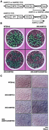
Figure 1.
Figure 1.
Establishment of the 35S:AtMYC2, 35S:AtMYB2, and 35S:AtMYC2/AtMYB2 Plants. (A) Scheme of the plant expression vectors. The constructs contain the 35S promoter of Cauliflower mosaic virus and Tobacco mosaic virus Ω sequence (Gallie et al., 1987) fused to AtMYC2 or AtMYB2 cDNAs. The AtMYC2 cDNA was cloned into the NotI site of the vector pBI121ΩKm or pBI121ΩHyg (AtMYC2/pBI121ΩKm or AtMYC2/pBI121ΩHyg), and AtMYB2 cDNA was cloned into the NotI site of the vector pBI121ΩKm (AtMYB2/pBI121ΩKm). Next, plasmids containing the AtMYC2 or AtMYB2 cDNA in the sense direction were identified by sequence analysis. AtMYC2/pBI121ΩKm and AtMYB2/pBI121ΩKm were used for the transformation of Arabidopsis. To make 35S:AtMYC2/AtMYB2 plants, 35S:AtMYB2 plants were retransformed by AtMYC2/pBI121ΩHyg. HPT, hygromycin phosphotransferase; NPTII, neomycin phosphotransferase II; Pnos and Tnos, nopaline synthase promoter and terminator, respectively. (B) Morphology of 35S:AtMYC2, 35S:AtMYB2, and 35S:AtMYC2/AtMYB2. Two-week-old seedlings of 35S:AtMYC2, 35S:AtMYB2, and 35S:AtMYC2/AtMYB2 grown axenically in biopots are shown. WT [Km] indicates the control plants transformed with the NPTII gene. (C) Paradermal micrographs of 35S:AtMYC2, 35S:AtMYB2, and 35S:AtMYC2/AtMYB2. Micrographs of 2-week-old plants were taken using Nomarski optics. All micrographs were taken at the same scale. Left panels represent epidermal cells, and right panels represent the palisade parenchyma. Bar = 100 μm.
Figure 2.
Figure 2.
Comparison of Growth Inhibition by ABA in 35S:AtMYC2, 35S:AtMYB2, and 35S:AtMYC2/AtMYB2 Plants. (A) Three-week-old seedlings of 35S:AtMYC2, 35S:AtMYB2, and 35S:AtMYC2/AtMYB2 grown on GM agar plates with or without 0.7 μM ABA. Transgenic plants with the NPTII gene (WT [Km]) were used as control wild-type plants. (B) Dry weight of 35S:AtMYC2, 35S:AtMYB2, and 35S:AtMYC2/AtMYB2 seedlings grown on GM agar plates containing ABA at 0, 300, 500, 700, or 1000 nM. The dry weight of each transgenic plant was measured at 30 days after the seeds were sown. Each value represents the average of >80 transformants with the standard error of at least two replicates. Transgenic plants with the NPTII gene (WT [Km]) and transgenic plants with the NPTII and HPT genes (WT [Km/Hyg]) were used as control wild-type plants.
Figure 3.
Figure 3.
Effect of the ABA Sensitivity of 35S:AtMYC2, 35S:AtMYB2, and 35S:AtMYC2/AtMYB2 Plants on Seed Germination Rate. The seed germination rates of 35S:AtMYC2, 35S:AtMYB2, and 35S:AtMYC2/AtMYB2 were measured on GM agar plates containing 0, 0.5, 1, 2, 3, or 5 μM ABA at 4 to 13 days after sowing. Each value is the average of >80 transformants with the standard error of at least two replicates. Transgenic plants with the NPTII gene (WT [Km]) and transgenic plants with the NPTII and HPT genes (WT [Km/Hyg]) were used as control wild-type plants. Percentages of germinated seeds were obtained and scored as germination rates.
Figure 4.
Figure 4.
RNA Gel Blot Analysis of the Expression of the rd22 and AtADH1 Genes. (A) and (B) To analyze the expression of the rd22 (A) and AtADH1 (B) genes, each lane was loaded with 30 μg of total RNA prepared from Arabidopsis plants (35S:AtMYC2, 35S:AtMYB2, 35S:AtMYC2/AtMYB2, and WT [Km]) that had been treated with ABA for 10 h at the indicated concentrations. The plants were transferred to water for 20 h and then treated with ABA. DNA fragments of the full-length rd22 and AtADH1 cDNAs were used as probes. EtBr, ethidium bromide. (C) and (D) To analyze the expression of AtMYC2 (C) and AtMYB2 (D), each lane was loaded with 30 μg of total RNAs prepared from untreated transgenic Arabidopsis plants. DNA fragments of the full-length AtMYC2 and AtMYB2 cDNAs were used as probes.
Figure 5.
Figure 5.
Comparison of the Stress Tolerance of 35S:AtMYC2/AtMYB2 and Wild-Type Plants Using Electrolyte Leakage. Three-week-old Arabidopsis rosette-stage plants were used to quantify electrolyte leakage. The plants were removed from the agar plates and grown hydroponically in water for 20 h (0 h control) and subsequently grown in mannitol solution at various concentrations (0, 400, 500, and 600 mM) for 4 h.
Figure 6.
Figure 6.
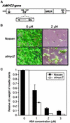
Disruption of the AtMYC2 Gene by Transposon-Mediated Mutagenesis Causes Insensitivity to ABA. (A) Scheme of the AtMYC2 gene showing the Ds insertion site between amino acids 107 and 108. The arrow indicates the direction of the Ds insertion. (B) Four-week-old atmyc2 mutant and Nossen wild-type Arabidopsis seedlings grown on GM agar plates with or without 2 μM ABA. (C) Dry weight of the atmyc2 mutant and wild-type Arabidopsis seedlings grown on GM agar plates containing 0, 1, 2, or 3 μM ABA. The dry weight of each transgenic plant was measured at 30 days after sowing. Each value represents the average of >80 transformants.
Figure 7.
Figure 7.
Effect of the ABA Sensitivity of the atmyc2 Mutant and Wild-Type Plants on Seed Germination Rate. The seed germination rates of atmyc2 and wild-type plants were measured on GM agar plates containing 0, 1, 2, or 3 μM ABA at 4 to 11 days after sowing. Each value represents the average of >80 transformants with the standard error of at least two replicates. Percentages of germinated seeds were obtained and scored as germination rates.
Figure 8.
Figure 8.
RNA Gel Blot Analysis of the Expression of the rd22, AtMYC2, and AtADH1 Genes in atmyc2 Mutant and Wild-Type Plants. (A) To analyze the expression of AtMYC2, each lane was loaded with 30 μg of total RNA prepared from atmyc2 mutant and Nossen wild-type plants (No-0) that had been treated for 5 h with 100 μM ABA. Cont., control treatment with water; EtBr, ethidium bromide. (B) and (C) To analyze the expression of the rd22 and AtADH1 genes, each lane was loaded with 30 μg of total RNAs prepared from atmyc2 mutant and Nossen plants that had been treated with ABA for 5 h at the designated concentrations. The plants were transferred to water for 20 h and then treated with ABA. DNA fragments of the full-length AtMYC2, rd22, and AtADH1 cDNAs were used as probes. (D) To quantify the expression of rd22 and AtADH1 genes, the intensity of each band was quantified by densitometry.
Figure 9.
Figure 9.
Phylogenic Tree of bHLH-Related Proteins and Comparison of Amino Acid Sequences of AtMYC2 and Its Homologs, AtMYC3 and AtMYC4. (A) The bootstrapped tree file was produced by CLUSTAL W from sequences of bHLH-related proteins. We searched the Arabidopsis genome database for amino acid sequences of the bHLH DNA binding domains of several bHLH-related proteins including AtMYC2 as a query. MIPS identifiers are shown by their protein names. The AtMYC2, AtMYC3, and AtMYC4 proteins are indicated in red. We basically selected the genes whose ESTs were obtained to construct the phylogenic tree except for At1g25330, At5g56960, At1g12540, At4g00120, At5g67060, and At1g27740. (B) The deduced amino acid sequence of AtMYC2 is compared with the sequences of AtMYC3 and AtMYC4. Asterisks represent identical amino acid residues, and dashes indicate gaps introduced to maximize alignment. The N-terminal unique domains of the bHLH family are highlighted in green, and N-terminal conserved regions and bHLH DNA binding domains are shown in red and purple. Triangles show repeated hydrophobic residues, which extend from helix II into the putative Leu zipper. (C) To analyze the expression of AtMYC2, AtMYC3, and AtMYC4, each lane was loaded with 30 μg of total RNA prepared from unbolted wild-type (Columbia) plants that had been dehydrated (Dry) and transferred from agar plates to hydroponic growth conditions in 100 μM ABA, 250 mM NaCl, or water. The plants were transferred to water for 20 h and then given various treatments. The DNA fragments of the full-length AtMYC2 and partial AtMYC3 and AtMYC4 cDNAs were used as probes. Numbers above each lane indicate the time in hours after the initiation of treatment.

References

    1. Abe, H., Yamaguchi-Shinozaki, K., Urao, T., Iwasaki, T., and Shinozaki, K. (1997). Role of MYC and MYB homologs in drought- and abscisic acid-regulated gene expression. Plant Cell 9, 1859–1868. - PMC - PubMed
    1. Bechtold, N., Ellis, J., and Pelletier, G. (1993). In planta Agrobacterium-mediated gene transfer by infiltration of adult Arabidopsis thaliana plants. C. R. Acad. Sci. Paris 316, 1194–1199.
    1. Becker, D. (1990). Binary vectors which allow the exchange of plant selectable markers and reporter genes. Nucleic Acids Res. 11, 203. - PMC - PubMed
    1. Bray, E.A. (1997). Plant responses to water deficit. Trends Plant Sci. 2, 48–54.
    1. Busk, P.K., and Pages, M. (1998). Regulation of abscisic acid-induced transcription. Plant Mol. Biol. 37, 425–435. - PubMed

Publication types

MeSH terms