. 2003 Jan;15(1):237-49.
doi: 10.1105/tpc.007153.

Actin-depolymerizing factor mediates Rac/Rop GTPase-regulated pollen tube growth

Affiliations

Actin-depolymerizing factor mediates Rac/Rop GTPase-regulated pollen tube growth

Christine Y-h Chen et al. Plant Cell. 2003 Jan.

Abstract

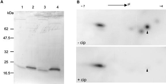
Pollen tube elongation is a rapid tip growth process that is driven by a dynamic actin cytoskeleton. A ubiquitous family of actin binding proteins, actin-depolymerizing factors (ADFs)/cofilins, bind to actin filaments, induce severing, enhance depolymerization from their slow-growing end, and are important for maintaining actin dynamics in vivo. ADFs/cofilins are regulated by multiple mechanisms, among which Rho small GTPase-activated phosphorylation at a terminal region Ser residue plays an important role in regulating their actin binding and depolymerizing activity, affecting actin reorganization. We have shown previously that a tobacco pollen-specific ADF, NtADF1, is important for maintaining normal pollen tube actin cytoskeleton organization and growth. Here, we show that tobacco pollen grains accumulate phosphorylated and nonphosphorylated forms of ADFs, suggesting that phosphorylation could be a regulatory mechanism for their activity. In plants, Rho-related Rac/Rop GTPases have been shown to be important regulators for pollen tube growth. Overexpression of Rac/Rop GTPases converts polar growth into isotropic growth, resulting in pollen tubes with ballooned tips and a disrupted actin cytoskeleton. Using the Rac/Rop GTPase-induced defective pollen tube phenotype as a functional assay, we show that overexpression of NtADF1 suppresses the ability of NtRac1, a tobacco Rac/Rop GTPase, to convert pollen tube tip growth to isotropic growth. This finding suggests that NtADF1 acts in a common pathway with NtRac1 to regulate pollen tube growth. A mutant form of NtADF1 with a nonphosphorylatable Ala substitution at its Ser-6 position [NtADF1(S6A)] shows increased activity, whereas the mutant NtADF1(S6D), which has a phospho-mimicking Asp substitution at the same position, shows reduced ability to counteract the effect of NtRac1. These observations suggest that phosphorylation at Ser-6 of NtADF1 could be important for its integration into the NtRac1 signaling pathway. Moreover, overexpression of NtRac1 diminishes the actin binding activity of green fluorescent protein (GFP)-NtADF1 but has little effect on the association of GFP-NtADF1(S6A) with actin cables in pollen tubes. Together, these observations suggest that NtRac1-activated activity regulates the actin binding and depolymerizing activity of NtADF1, probably via phosphorylation at Ser-6. This notion is further supported by the observation that overexpressing a constitutively active NtRac1 in transformed pollen grains significantly increases the ratio of phosphorylated to nonphosphorylated ADFs. Together, the observations reported here strongly support the idea that NtRac1 modulates NtADF1 activity through phosphorylation at Ser-6 to regulate actin dynamics.


Figures

Figure 1.
Figure 1.
Tobacco Pollen ADFs Are Phosphorylated. (A) An immunoblot of lily (lanes 1 and 2) and tobacco (lanes 3 and 4) pollen grain proteins after SDS-PAGE (17.5%). The blot was reacted with anti-NtADF1 antibodies and alkaline phosphatase–conjugated secondary antibodies. The molecular mass of the detected protein bands was ∼17 kD, approximating the predicted molecular mass of pollen ADFs from these species (Kim et al., 1993; Chen et al., 2002). Lanes 2 and 4 contained fourfold concentrated versions of the samples loaded in lanes 1 and 3, respectively. The slightly retarded mobility of the NtADF protein bands in lanes 2 and 4 probably resulted from slightly higher salt concentrations in these concentrated protein samples. (B) Immunoblots of tobacco pollen proteins after two-dimensional SDS-PAGE (17.5%). Both blots were reacted with anti-NtADF1 antibodies and horseradish peroxidase–conjugated secondary antibodies. NtADF spots were revealed by chemiluminescence detection. Both gels contained proteins that were not (−cip) or were (+cip) treated with alkaline phosphatase. Approximately 60 μg of total pollen protein was loaded in the −cip sample. The same amount of protein was treated with alkaline phosphatase in the +cip sample. The amount of protein loaded in the +cip sample was adjusted so that the intensity of the more alkaline NtADF species in the −cip and +cip samples was comparable to highlight the difference in the intensity of the more acidic NtADFs. Arrowheads point to the location of the more acidic NtADF spot. The minor protein spots detected by NtADF1 antibodies at the acidic range of the blot may represent minor pollen-expressed NtADFs.
Figure 2.
Figure 2.
NtRac1 Induces Ectopic Pollen Tube Expansion and the Disruption of Normal Actin Organization. (A) Epifluorescent micrographs of the GFP-NtRac1–induced pollen tube phenotype. The top panel shows a pollen tube that exhibited a balloon-tipped phenotype soon after germination. The other panels show pollen tubes that had elongated for various times before terminating in a balloon-tipped phenotype. Pollen tubes similar to those shown in the top two panels are most characteristic of those that have ballooned at early time points (3 to 4 h) after germination. The pollen tubes shown in the third and fourth panels are characteristic of those that have elongated farther and for longer periods of time before balloon formation and growth arrest. Arrowheads indicate the ballooned tip region. All images were taken by exposure time automatically set by the camera. In general, pollen tubes that ballooned early were more fluorescent than those that maintained a normal morphology at the same time point, suggesting a correlation of the phenotype with the level of GFP-NtRac1 expression (Cheung et al., 2003). Cotransformation using Lat52-GFP as a marker gene for transformation and Lat52-NtRac1 as the regulator gene resulted in qualitatively similar observations except that the more severe phenotypes, such as those shown in the top two panels, predominated even at the earliest time point. Bars = 10 μm. (B) Confocal images of two pollen tubes transformed by Lat52-GFP-mTalin. GFP-mTalin revealed long actin cables aligned along the long axis of the pollen tube shank, which is characteristic of elongating pollen tubes (Kost et al., 1998, 1999; Vidali et al., 2001; Chen et al., 2002). The tip of the pollen tube shown in the top panel pointed upward slightly, leading to the impression of a bulge at the tip when sections across the entire tubes were projected. The actin filaments seen at the center of the bulge represent fine actin filaments close to the cortical region of the tube tip. Typically, long actin cables do not invade the apical region, as shown in the bottom panel. The pollen tubes shown here had the best tube morphology and actin cytoskeleton structure among transformed pollen tubes that expressed levels of GFP-mTalin detectable with our confocal microscope. (C) and (D) Confocal images of pollen tubes cotransformed by Lat52-NtRac1 and Lat52-GFP-mTalin to visualize the actin cytoskeleton in tubes affected by the overexpression of NtRac1. (C) This pollen tube was converting from tip growth to isotropic growth. The shank actin cables still were aligned axially, but they started to extend farther into the tip region than in normally tip-elongating tubes. (D) A balloon-tipped pollen tube showing highly bundled actin cables in the shank and an extensive network of intertwining actin cables in the ballooned tip. Bars in (B) to (D) = 10 μm.
Figure 3.
Figure 3.
NtADF1 Counteracts the Ability of NtRac1 to Induce Isotropic Pollen Tube Growth, and Ser-6 in NtADF1 Is Important for This Activity. Pollen grains were cotransformed with Lat52-GFP-NtRac1 (A) or Lat52-GFP-NtRac1(G15V) and Lat52-NtADF1, Lat52-NtADF1(S6A), or Lat52-NtADF(S6D), as indicated (B). Lat52-GUS was included in the control samples to ensure that comparable amounts of DNA were introduced into the transformed pollen grains. Pollen tubes were scored at 3 h after germination. Pollen tube tips that were equal to or larger than the diameter of the pollen grains were scored as balloon tipped. Data were obtained from three independent experiments. In each experiment, every condition was tested three times. The total number of pollen tubes scored (n) for each condition is indicated above the data bars. Error bars indicate standard deviations. t test analysis of the data shown in (B) indicates the level of significance to be P = 0.000023, 0.000016, and 0.499 for the NtADF1, NtADF1(S6A), and NtADF1(S6D) data, respectively, relative to the control data. The level of significance between the NtADF1 data and the NtADF1(S6A) and NtADF1(S6D) data was P = 0.08588 and 0.00013, respectively.
Figure 4.
Figure 4.
NtRac1 Reduces Interactions between NtADF1 and Actin in Transformed Pollen Tubes. The recombinant protein expressed in each of the transformed pollen tubes shown is indicated. (A) A normally elongating transformed pollen tube showing the GFP-NtADF1 association with long actin cables in the shank and an actin mesh at the subapical region, as reported previously (Chen et al., 2002). (B) A balloon-tipped pollen tube that was cotransformed with Lat52-NtRac1 and Lat52-GFP-NtADF1 showing green fluorescence through-out the tube but no discrete association with filamentous structures. By contrast, some thin GFP-labeled actin cables were observed amid a strong cytosolic green fluorescent signal, reflecting the effect of the overexpressed NtRac1, in tubes that still maintained the normal tube morphology at the same time point (data not shown). (C) A balloon-tipped pollen tube that was transformed with Lat52-NtRac1 and Lat52-GFP-NtADF1(S6A) showing that the association between the GFP-tagged mutant NtADF1 and the actin cables was preserved, indicating that the actin binding activity of GFP-NtADF1(S6A) is resistant to the effect of NtRac1. (D) A highly disorganized actin network in the ballooned tip region, similar to but less bundled than the GFP-mTalin, revealed actin cytoskeleton in NtRac1-coexpressing balloon-tipped pollen tubes.
Figure 5.
Figure 5.
Overexpressing NtRac1(G15V) Increases the Ratio of Phosphorylated to Nonphosphorylated Forms of Pollen ADF. (A) Immunoblot of proteins from pollen grains isolated from a representative Lat52-NtRac1(G15V)–transformed plant after two-dimensional SDS-PAGE. Densitometric scanning of immunoblots prepared from three different preparations of pollen proteins indicates a ratio of at least 3.5 for the phosphorylated ADF to the nonphosphorylated form, more than a threefold increase from that observed among ADFs from control wild-type pollen. Approximately 60 μg of pollen proteins was loaded. (B) Germination cultures of pollen grains from a representative Lat52-NtRac1(G15V)–transformed plant. The arrow in the inset points to the expanded tip of a just-emerged pollen tube, which is characteristic of most of the germinating pollen grains.
Figure 6.
Figure 6.
A Model for Rac/Rop GTPase Signaling to the Pollen Tube Actin Cytoskeleton via ADF. The cycling of actin polymerization and depolymerization is shown in the middle of the figure. Arrow blocks with T or D embedded within represent actin monomers. T indicates ATP-bound, D indicates ADP-bound, and D-pi indicates ADF- and Pi-bound actin monomers (Bamburg, 1999). The minus (pointed) and plus (barbed) ends of the actin filaments are shown accordingly. The equilibrium of nonphosphorylated ADF and phosphorylated ADF (pADF) and their activity is depicted as regulated by Rac/Rop GTPase–activated, yet to be identified enzyme(s). ADF dephosphorylation and its regulatory mechanisms also remain to be revealed. The possible effect of Rac/Rop-regulated phospholipid PIP and PIP2 conditions on ADF activity is indicated by the long dotted arrow. The phosphoinositide-mediated changes in intracellular Ca2+ conditions also may affect the phosphorylation or dephosphorylation of ADFs (indicated by the short dotted arrow) if the enzymes involved are regulated by this ion. Active ADFs bind to actin and have higher affinity for ADP actin; thus, they preferentially bind to and enhance depolymerization from the pointed end (Carlier et al., 1997; Bamburg, 1999). ADF also binds to actin cooperatively, so multiple ADF molecules may be bound to an actin filament, inducing filament severing (McGough et al., 1997; McGough and Chiu, 1999). pADF does not associate with actin cables. The typical actin cytoskeleton structure as revealed by GFP-NtADF1 labeling in elongating pollen tubes (Chen et al., 2002) is shown at bottom. The level of actin cycling that maintains the actin cytoskeleton organization in elongating pollen tubes would depend on the combined activities from regulated levels of ADF, pADF, and other actin binding proteins to affect polar tube growth (Hepler et al., 2001).

Similar articles

Cited by

References

    1. Agnew, B.J., Minamide, L.S., and Bamburg, J.R. (1995). Reactivation of phosphorylated actin depolymerizing factor and identification of the regulatory site. J. Biol. Chem. 270, 17582–17587. - PubMed
    1. Allwood, E.G., Smertenko, A.P., and Hussey, P.J. (2001). Phosphorylation of plant actin-depolymerizing factor by calmodulin-like domain protein kinase. FEBS Lett. 499, 97–100. - PubMed
    1. Arabidopsis Genome Initiative (2000). Analysis of the genome sequence of the flowering plant Arabidopsis thaliana. Nature 408, 796–815. - PubMed
    1. Arber, S., Barbayannis, F.A., Hanser, H., Schindler, C., Stanton, C.A., Bernard, O., and Caroni, P. (1998). Regulation of actin dynamics through phosphorylation of cofilin by LIM-kinase. Nature 393, 805–809. - PubMed
    1. Ausubel, F.M., Brent, R., Kingston, R.E., Moore, D.D., Seidman, J.G., Smith, J.A., and Struhl, K. (2000). Current Protocols in Molecular Biology. (New York: John Wiley & Sons).

Publication types

MeSH terms

Associated data