

Cholecystokinin-like immunoreactive amacrine cells in the rat retina
- PMID: 12511085
- PMCID: PMC3342674
- DOI: 10.1017/s0952523802194156
Cholecystokinin-like immunoreactive amacrine cells in the rat retina
Abstract
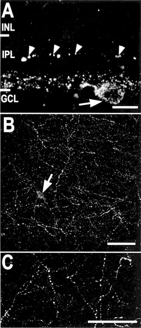
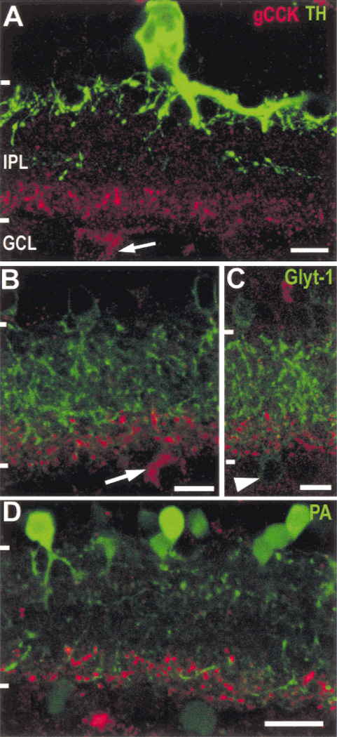
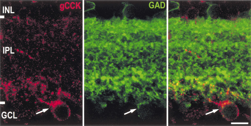
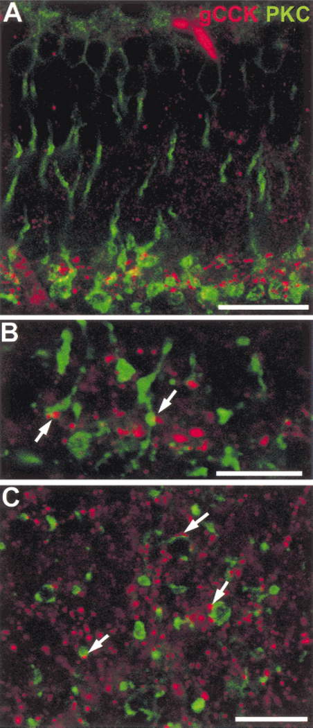
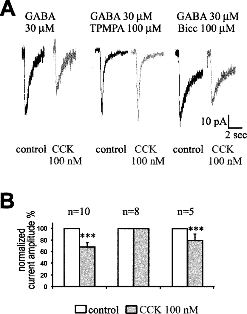
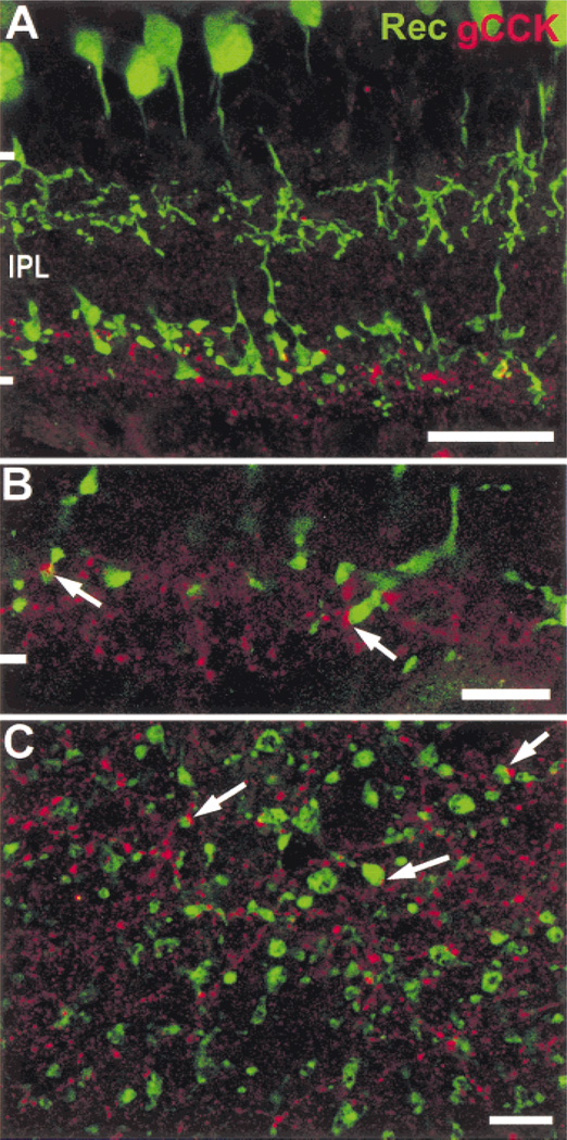
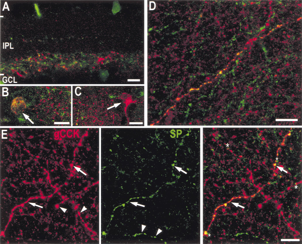
High levels of endogenous cholecystokinin (CCK) are present in the rat retina (Eskay & Beinfeld, 1982), but the cellular localization and physiological actions of CCK in the rat retina are uncertain. The goals of this study were to characterize the cells containing CCK, identify cell types that interact with CCK cells, and investigate the effects of CCK on rod bipolar cells. Rat retinas were labeled with antibody to gastrin-CCK (gCCK) using standard immunofluorescence techniques. Patch-clamp methods were used to record from dissociated rod bipolar cells from rats and mice. Gastrin-CCK immunoreactive (-IR) axons were evenly distributed throughout the retina in stratum 5 of the inner plexiform layer of the rat retina. However, the gCCK-IR somata were only detected in the ganglion cell layer in the peripheral retina. The gCCK-IR cells contained glutamate decarboxylase, and some of them also contained immunoreactive substance P. Labeled axons contacted PKC-IR rod bipolar cells, and recoverin-IR ON-cone bipolar cells. CCK-octapeptide inhibits GABA(C) but not GABA(A) mediated currents in dissociated rod bipolar cells.
Figures
Similar articles
-
The neurons of the ground squirrel retina as revealed by immunostains for calcium binding proteins and neurotransmitters.J Neurocytol. 2002 Sep-Nov;31(8-9):649-66. doi: 10.1023/a:1025791512555. J Neurocytol. 2002. PMID: 14501205
-
Depletion of cholinergic amacrine cells by a novel immunotoxin does not perturb the formation of segregated on and off cone bipolar cell projections.J Neurosci. 2002 Mar 15;22(6):2265-73. doi: 10.1523/JNEUROSCI.22-06-02265.2002. J Neurosci. 2002. PMID: 11896166 Free PMC article.
-
Recoverin immunoreactivity in mammalian cone bipolar cells.Vis Neurosci. 1993 Jan-Feb;10(1):1-12. doi: 10.1017/s0952523800003175. Vis Neurosci. 1993. PMID: 8424920
-
Ectopic photoreceptors and cone bipolar cells in the developing and mature retina.J Neurosci. 2003 Feb 15;23(4):1383-9. doi: 10.1523/JNEUROSCI.23-04-01383.2003. J Neurosci. 2003. PMID: 12598626 Free PMC article.
-
Goalpha labels ON bipolar cells in the tiger salamander retina.J Comp Neurol. 2003 Jun 23;461(2):276-89. doi: 10.1002/cne.10704. J Comp Neurol. 2003. PMID: 12724843
Cited by
-
Cholecystokinin receptor type A are involved in the circadian rhythm of the mouse retina.Heliyon. 2024 Jun 7;10(12):e32653. doi: 10.1016/j.heliyon.2024.e32653. eCollection 2024 Jun 30. Heliyon. 2024. PMID: 39183886 Free PMC article.
-
Comprehensive single-cell atlas of the mouse retina.bioRxiv [Preprint]. 2024 Jan 28:2024.01.24.577060. doi: 10.1101/2024.01.24.577060. bioRxiv. 2024. Update in: iScience. 2024 May 08;27(6):109916. doi: 10.1016/j.isci.2024.109916. PMID: 38328114 Free PMC article. Updated. Preprint.
-
Mouse Retinal Cell Atlas: Molecular Identification of over Sixty Amacrine Cell Types.J Neurosci. 2020 Jul 1;40(27):5177-5195. doi: 10.1523/JNEUROSCI.0471-20.2020. Epub 2020 May 26. J Neurosci. 2020. PMID: 32457074 Free PMC article.
-
All spiking, sustained ON displaced amacrine cells receive gap-junction input from melanopsin ganglion cells.Curr Biol. 2015 Nov 2;25(21):2763-2773. doi: 10.1016/j.cub.2015.09.018. Epub 2015 Oct 1. Curr Biol. 2015. PMID: 26441349 Free PMC article.
-
Comprehensive single-cell atlas of the mouse retina.iScience. 2024 May 8;27(6):109916. doi: 10.1016/j.isci.2024.109916. eCollection 2024 Jun 21. iScience. 2024. PMID: 38812536 Free PMC article.
References
-
- Ayoub GS, Matthews G. Substance P modulates calcium current in retinal bipolar neurons. Visual Neuroscience. 1992;8:539–544. - PubMed
-
- Beinfeld MC. Cholecystokinin/Gastrin. In: Watson SJ, editor. Psychopharmacology. New York: Lippincott-Raven Press; 1998. CD/ROM Edition http://www.acnp.org/citations/ GN401000056: Chapter 56.
-
- Blanco R, Vaquero CF, De La Villa P. Action potentials in axonless horizontal cells isolated from the rabbit retina. Neuroscience Letters. 1996;203:57–60. - PubMed
-
- Bone EA, Rosenzweig SA. Characterization of cholecystokinin receptors in toad retina. Peptides. 1988;9:373–381. - PubMed
-
- Brandon C. Retinal GABA neurons: Localization in vertebrate species using an antiserum to rabbit brain glutamate decarboxylase. Brain Research. 1985;344:286–295. - PubMed
Publication types
MeSH terms
Substances
Grants and funding
LinkOut - more resources
Full Text Sources
Miscellaneous
