. 2003 Jan;185(2):413-21.
doi: 10.1128/JB.185.2.413-421.2003.

Transcriptional regulation in the Streptococcus pneumoniae rlrA pathogenicity islet by RlrA

Affiliations

Transcriptional regulation in the Streptococcus pneumoniae rlrA pathogenicity islet by RlrA

David L Hava et al. J Bacteriol. 2003 Jan.

Abstract

The proper temporal expression of virulence genes during infection is crucial to the infectious life cycle of microbial pathogens, particularly in pathogens that encounter a multitude of environments in eukaryotic hosts. Streptococcus pneumoniae normally colonizes the nasopharynges of healthy adults but can cause a range of diseases at a variety of host sites. Transcriptional regulators that are essential for full virulence of S. pneumoniae in different animal models have been identified. One such regulator, rlrA, is required for colonization of the nasopharynx and lung infection but is dispensable for systemic infection. Previous work has shown that rlrA lies in a 12-kb pathogenicity islet, divergently opposed to three putative sortase-anchored surface proteins and three sortase enzymes. In addition to rlrA, one of the putative surface proteins and one of the sortases have also been shown to be essential for lung infection. In this work, we demonstrate that RlrA is a positive regulator of all seven genes in the rlrA pathogenicity islet, with transcriptional activation occurring at four different promoters in the islet with AT-rich sequences. These promoters direct the expression of rlrA itself, the three sortases, rrgA, and rrgBC. These data are consistent with the model whereby the rlrA pathogenicity islet acts in an autonomous manner to alter the bacterial surface components that interact with the pulmonary and nasopharyngeal environments.


Figures

FIG. 1.
FIG. 1.
The rlrA pathogenicity islet. The 12-kb locus includes a positive regulator, three surface proteins, and three sortase homologues. The four genes that are required for virulence in one or more animal models are in white (10).
FIG. 2.
FIG. 2.
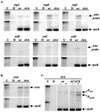
RPAs were performed to analyze the steady-state mRNA levels of each gene in the rlrA pathogenicity islet in both the wild-type (wt; AC353) and rlrA mutant (AC1213) strain backgrounds. (A) Riboprobes to each gene in the islet, as well as to rpoB, were generated and hybridized to 10 μg of total S. pneumoniae RNA from either the wild-type or the mutant strain. Lanes U and D contained undigested riboprobes or riboprobes digested by RNase in the absence of S. pneumoniae RNA. In each case, the experimental probe for the given gene in the upper part of the panel and the control rpoB probe in the lower part of the panel are from the same gel. (B) Riboprobes to srtA and rpoB were hybridized to the same samples in panel A and presented in the same manner. (C) A riboprobe that differentially recognizes the two rlrA transcripts in AC1278 was used to determine if RlrA is autoregulatory. The larger fragment in each lane represents the mRNA from the native rlrA pathogenicity islet promoter. Lanes marked with a plus sign are RNA samples that were harvested from cells grown in the presence of maltose.
FIG. 3.
FIG. 3.
(A) Transcriptional start sites of promoters upstream of rlrA, rrgA, rrgB, and srtB were mapped by primer extension analysis. The arrows indicate primer extension products. (B) Graphical depiction of the four rlrA pathogenicity islet promoters. A rightward arrow indicates the +1 start site. When present, −10 and −35 σ70 consensus sequences and predicted Shine-Dalgarno (SD) sequences are underlined and in bold.
FIG. 4.
FIG. 4.
Northern blot of rlrA pathogenicity islet mRNAs. Riboprobes to selected genes were synthesized and used to hybridize to total RNA recovered from AC1278 (lane 1) or AC1213 (lane 2) grown under maltose-inducing conditions. (A) Northern blots probed with rrgB and rrgC riboprobes. (B) Northern blots probed with srtB, srtC, and srtD riboprobes.
FIG. 5.
FIG. 5.
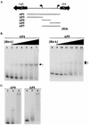
Gel mobility shift analysis with RlrA-His6. (A) The four 32P-labeled probes that span the rrgA-rlrA intergenic region and were used in gel mobility shift analyses are depicted. The sizes of the PCR fragments were as follows: AP1, 522 bp; AP3, 250 bp; AP4, 139 bp; AP5, 163 bp; AP7, 290 bp. (B) Gel mobility shift analysis of AP4 and AP5. 32P-labeled probes were incubated with increasing concentrations of RlrA-His6. The protein concentrations used were as follows: lanes 1 and 8, 0 nM; lanes 2 and 9, 0.25 nM; lanes 3 and 10, 1 nM; lanes 4 and 11, 4 nM; lanes 5 and 12, 16.4 nM; lanes 6 and 13, 33 nM; lanes 7 and 14, 66 nM. The arrows indicate shifted species. (C) Supershift of RlrA-His6 complexes by the addition of anti-His6 antibody to the binding reaction mixture. The protein concentrations used were as follows: lanes 1 and 4, no protein; lanes 2 and 5, 16.4 nM RlrA-His6; lanes 3 and 6, 16.4 nM RlrA-His6 and 0.5 μg of anti-His6 antibody.
FIG. 6.
FIG. 6.
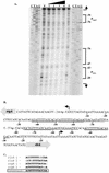
(A) DNase I footprinting analysis of the rrgA-rlrA promoter regions. The 32P-labeled AP7 probe was incubated with increasing amounts of RlrA-His6 and subsequently treated with DNase I. The protein concentration used were as follows: lane 1, 0 nM; lane 2, 0.5 nM; lane 3, 2.05 nM; lane 4, 8.2 nM; lane 5, 32.8 nM. The amounts of DNase I used were as follows: lanes 1 and 2, 0.5 U; lane 3, 1 U; lanes 4 and 5, 2 U. Brackets indicate areas protected by RlrA-His6. (B) The rlrA and rrgA promoter regions. The nucleotide sequence of each promoter is shown, and a hooked arrow denotes the +1 transcription initiation site. RlrA binding sites are singly underlined, and consensus binding sites are doubly underlined. (C) Alignment of the four consensus RlrA binding sites within the rlrA and rrgA promoter regions. Nucleotides that are identical to those in the consensus sequence are in bold, and the consensus sequence is in bold and boxed. In both cases, the sequence labeled number 1 is the consensus site closest to the transcriptional start site for that gene.

References

    1. Acebo, P., C. Nieto, M. A. Corrales, M. Espinosa, and P. Lopez. 2000. Quantitative detection of Streptococcus pneumoniae cells harbouring single or multiple copies of the gene encoding the green fluorescent protein. Microbiology 146:1267-1273. - PubMed
    1. Charpentier, E., and E. Tuomanen. 2000. Mechanisms of antibiotic resistance and tolerance in Streptococcus pneumoniae. Microbes Infect. 2:1855-1864. - PubMed
    1. Claverys, J. P., A. Dintilhac, E. V. Pestova, B. Martin, and D. A. Morrison. 1995. Construction and evaluation of new drug-resistance cassettes for gene disruption mutagenesis in Streptococcus pneumoniae, using an ami test platform. Gene 164:123-128. - PubMed
    1. Crook, D. W., and B. G. Spratt. 1998. Multiple antibiotic resistance in Streptococcus pneumoniae. Br. Med. Bull. 54:595-610. - PubMed
    1. Fogg, G. C., and M. G. Caparon. 1997. Constitutive expression of fibronectin binding in Streptococcus pyogenes as a result of anaerobic activation of rofA. J. Bacteriol. 179:6172-6180. - PMC - PubMed

Publication types

Substances

LinkOut - more resources