. 2003 Jan;111(1):71-9.
doi: 10.1172/JCI16157.

Distinct progenitor populations in skeletal muscle are bone marrow derived and exhibit different cell fates during vascular regeneration

Affiliations

Distinct progenitor populations in skeletal muscle are bone marrow derived and exhibit different cell fates during vascular regeneration

Susan M Majka et al. J Clin Invest. 2003 Jan.

Abstract

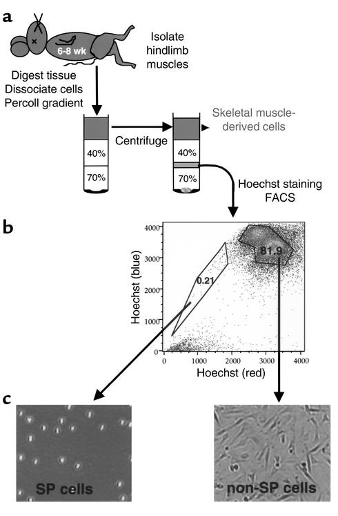
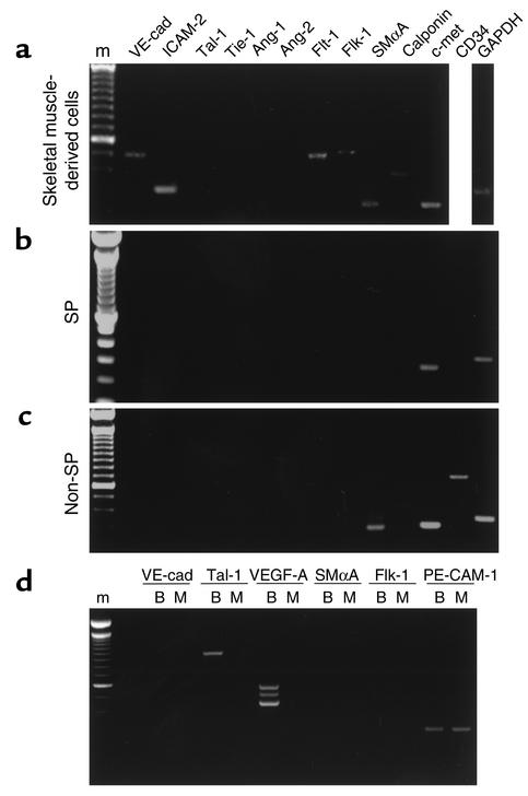
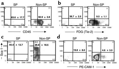
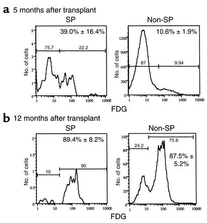
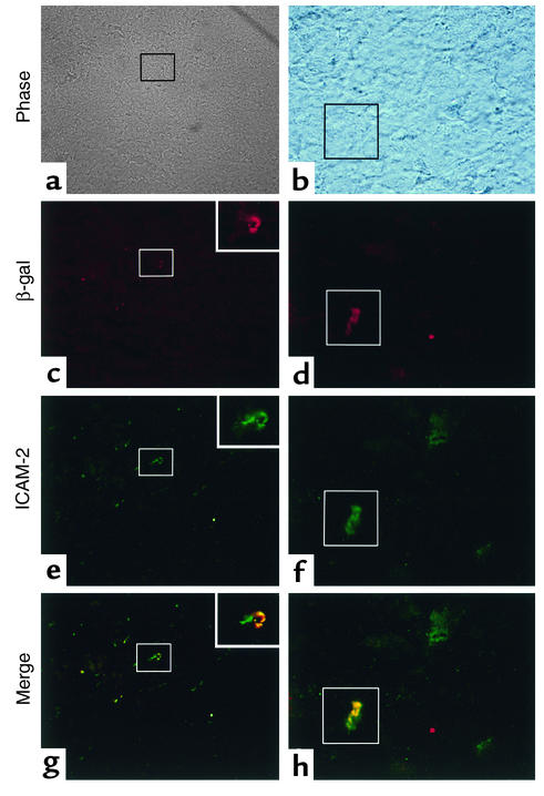
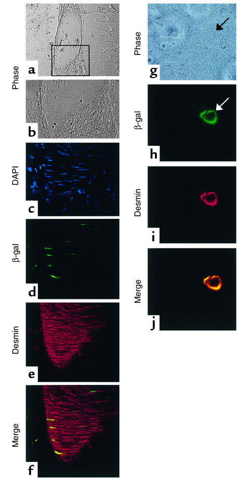
Vascular progenitors were previously isolated from blood and bone marrow; herein, we define the presence, phenotype, potential, and origin of vascular progenitors resident within adult skeletal muscle. Two distinct populations of cells were simultaneously isolated from hindlimb muscle: the side population (SP) of highly purified hematopoietic stem cells and non-SP cells, which do not reconstitute blood. Muscle SP cells were found to be derived from, and replenished by, bone marrow SP cells; however, within the muscle environment, they were phenotypically distinct from marrow SP cells. Non-SP cells were also derived from marrow stem cells and contained progenitors with a mesenchymal phenotype. Muscle SP and non-SP cells were isolated from Rosa26 mice and directly injected into injured muscle of genetically matched recipients. SP cells engrafted into endothelium during vascular regeneration, and non-SP cells engrafted into smooth muscle. Thus, distinct populations of vascular progenitors are resident within skeletal muscle, are derived from bone marrow, and exhibit different cell fates during injury-induced vascular regeneration.


Figures

Figure 1
Figure 1
Isolation of murine skeletal muscle progenitors. (a) Skeletal muscle stem cells were isolated from 6- to 8-week-old C57Bl/6 or Rosa26 mice, fractionated on a Percoll gradient, stained with Hoechst 33342 dye, and subjected to FACS. SP and non-SP cells were clearly discernable based on Hoechst dye exclusion, and the distinct populations were isolated via FACS. (b) In this representative sample, the SP population was 0.21%, and the non-SP was 81.9% of the total viable cells. (c) Both SP and non-SP populations were cultured overnight, and their distinct morphologies were revealed upon microscopic examination of the cells in culture. The SP cells were found to be of uniform morphology and nonadherent. The non-SP population contained predominantly adherent, spindle-shaped cells.
Figure 2
Figure 2
Analysis of vascular-specific gene expression in skeletal muscle-derived cells. RT-PCR analysis was performed on total RNA (50 ng/sample) isolated from distinct populations of skeletal muscle cells: (a) Percoll-fractionated skeletal muscle cells; (b) muscle SP population; and (c) muscle non-SP population. (d) Bone marrow-derived SP cells were directly compared to skeletal muscle-derived SP cells. GAPDH was used as a loading control. Analyses using each primer set were performed on three to five independent RNA isolates from each cell population; representative data are shown. The amplicon generated from each primer set was cloned and sequenced to verify that the primers amplify the intended gene. VE-cad, VE-cadherin; SMαA, SM-α-actin; m, marker; B, bone marrow SP; M, muscle SP.
Figure 3
Figure 3
FACS analysis for vascular lineage markers in muscle SP and non-SP populations. FACS data were compiled from four experiments, and the expression of specific proteins, displayed as mean ± SD from these independent experiments, are shown: (a) CD45; (b) Tie-2, as evidenced by FDG cleavage, indicating β-gal activity driven from the Tie-2 promoter; (c) Sca-1; and (d) PE-CAM-1.
Figure 4
Figure 4
Vascular progenitors within skeletal muscle are derived from bone marrow. SP cells were isolated from bone marrow of Rosa26 mice and stably engrafted into lethally irradiated, genetically matched recipients. After 5 or 12 months, the hindlimb musculature was excised and examined for the presence of LacZ-positive vascular progenitors. Percoll-fractionated muscle cells were stained with Hoechst dye and loaded with FDG, then subjected to FACS to isolate muscle SP and non-SP populations and determine whether they exhibit β-gal activity. (a) The mean ± SD of LacZ-positive SP and non-SP cells among three mice at 5 months after transplant was 39.0% ± 16.4% and 10.6% ± 1.9%, respectively. (b) At 12 months after transplantation, the mean ± SD of LacZ-positive SP and non-SP cells among three mice was 89.4% ± 8.2% and 87.5% ± 5.2%, respectively.
Figure 5
Figure 5
Skeletal muscle-derived SP cells engraft into vascular endothelium. Six- to eight-week-old C57Bl/6 mice were anesthetized with Avertin, and both TA muscles were injected with 25 μl of a 1 mg/ml cardiotoxin. After 8 or 24 hours, the right limb TA muscle was injected with 2,000 LacZ-positive muscle SP cells, and the left leg was injected with HBSS alone. After 4 weeks, the TA muscles of each of four experimental animals were harvested and processed for frozen sectioning and immunohistochemical analysis. All sections were immunostained for β-gal and costained for either ICAM-2 or desmin to detect injury-induced engraftment of LacZ-positive SP cells into regenerated vascular endothelium and smooth muscle, respectively. β-gal expression in vascular structures was colocalized only with ICAM-2: (a and b) bright field, boxes indicate costained vessels; (c and d) β-gal immunohistochemistry; (e and f) ICAM-2 immunohistochemistry; (g and h) β-gal and ICAM-2 fluorescent images were merged to reveal areas of costaining, as evidenced by the yellow staining pattern. Magnification, ×400 (a, c, e, g); ×1,000 (b, d, f, h, and inset boxes in c, e, and g).
Figure 6
Figure 6
Skeletal muscle-derived non-SP cells engraft into vascular smooth muscle. Six- to eight- week-old C57Bl/6 mice were anesthetized with Avertin, and both TA muscles were injected with 25 μl of a 1 mg/ml cardiotoxin. After 8 or 24 hours, the right limb TA muscle was injected with 500,000 LacZ-positive muscle non-SP cells, and the left leg was injected with HBSS alone. After 4 weeks, the TA muscles of each of four experimental animals were harvested and processed for frozen sectioning and immunohistochemical analysis. All sections were immunostained for β-gal, and costained for either ICAM-2 or desmin to detect injury-induced engraftment of LacZ positive non-SP cells into regenerated vascular endothelium and smooth muscle, respectively. β-gal expression in vascular structures colocalized only with desmin: (a, b, and g) bright field, box in a indicates region shown in (bf); (c) DAPI; (d and h) β-gal immunohistochemistry; (e and i) desmin immunohistochemistry; (f and j) β-gal and desmin fluorescent images were merged to reveal areas of costaining, as evidenced by the yellow staining pattern. Magnification, ×200 (a); ×600 (b, c, d, e, and f); ×1,000 (g, h, i, and j).

Comment in

  • Machinations of the marrow.
    Rosenthal N. Rosenthal N. J Clin Invest. 2003 Jan;111(1):29-30. doi: 10.1172/JCI17546. J Clin Invest. 2003. PMID: 12511584 Free PMC article. Review. No abstract available.

References

    1. Asahara T, et al. Isolation of putative progenitor endothelial cells for angiogenesis. Science. 1997;275:964–967. - PubMed
    1. Schatteman GC, et al. Blood-derived angioblasts accelerate blood flow restoration in diabetic mice. J. Clin. Invest. 2000;106:571–578. - PMC - PubMed
    1. Shimizu K, et al. Host bone marrow cells are a source of donor intimal smooth muscle-like cells in murine aortic transplant arteriopathy. Nat. Med. 2001;7:738–741. - PubMed
    1. Jackson KA, et al. Regeneration of cardiac muscle and vascular endothelium by adult stem cells. J. Clin. Invest. 2001;107:1395–1402. - PMC - PubMed
    1. Orlic D, et al. Bone marrow cells regenerate infarcted myocardium. Nature. 2001;410:701–705. - PubMed

Publication types