. 2003 Jan;69(1):56-65.
doi: 10.1128/AEM.69.1.56-65.2003.

ACEI of Trichoderma reesei is a repressor of cellulase and xylanase expression

Affiliations

ACEI of Trichoderma reesei is a repressor of cellulase and xylanase expression

Nina Aro et al. Appl Environ Microbiol. 2003 Jan.

Abstract

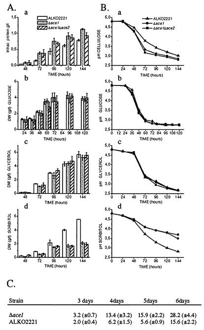
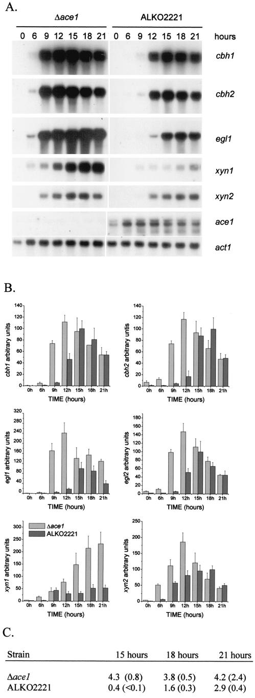
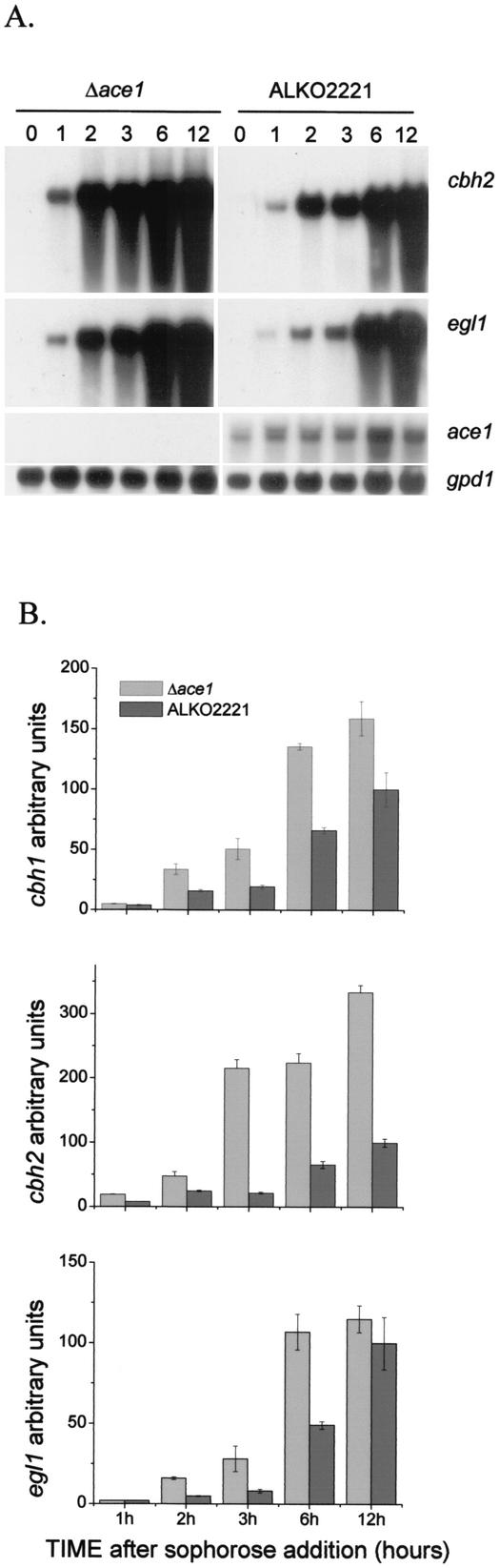
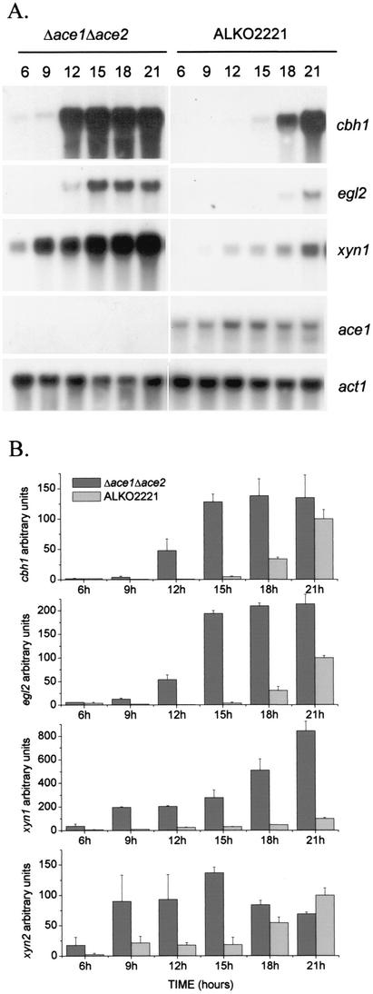
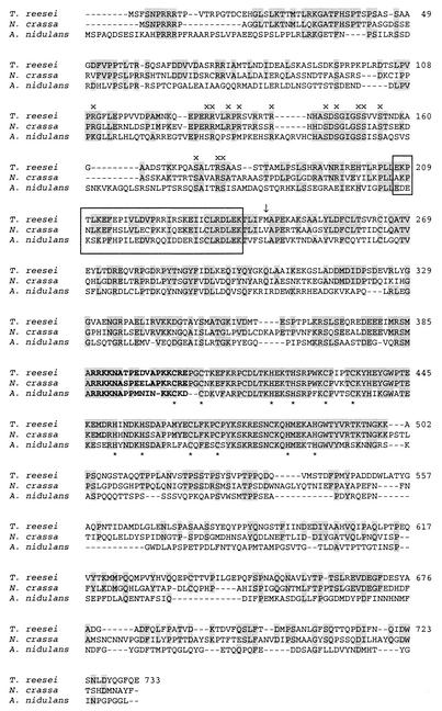
We characterized the effect of deletion of the Trichoderma reesei (Hypocrea jecorina) ace1 gene encoding the novel cellulase regulator ACEI that was isolated based on its ability to bind to and activate in vivo in Saccharomyces cerevisiae the promoter of the main cellulase gene, cbh1. Deletion of ace1 resulted in an increase in the expression of all the main cellulase genes and two xylanase genes in sophorose- and cellulose-induced cultures, indicating that ACEI acts as a repressor of cellulase and xylanase expression. Growth of the strain with a deletion of the ace1 gene on different carbon sources was analyzed. On cellulose-based medium, on which cellulases are needed for growth, the Deltaace1 strain grew better than the host strain due to the increased cellulase production. On culture media containing sorbitol as the sole carbon source, the growth of the strain with a deletion of the ace1 gene was severely impaired, suggesting that ACEI regulates expression of other genes in addition to cellulase and xylanase genes. A strain with a deletion of the ace1 gene and with a deletion of the ace2 gene coding for the cellulase and xylanase activator ACEII expressed cellulases and xylanases similar to the Deltaace1 strain, indicating that yet another activator regulating cellulase and xylanase promoters was present.


Figures

FIG. 1.
FIG. 1.
(A and B) Growth of the ALKO2221, Δace1 (VTT-D-01850 and VTT-D-01849), and Δace1 Δace2 (VTT-D-01851 and VTT-D-01852) strains on cellulose (panels a), glucose (panels b), glycerol (panels c), and sorbitol (panels d). (A) Mycelial biomasses or total amounts of intracellular protein of the ALKO2221, Δace1, and Δace1 Δace2 strains at different times. The error bars indicate the standard deviations of the mean dry weights (DW) and amounts of total protein. (B) pH values of the culture media of the ALKO2221, Δace1, and Δace1 Δace2 strains. (C) Cellulase activities (in nanokatals per milliliter; 1 nkat =1 mmol of methylumbelliferyl released from MUL) in cellulose culture supernatants of Δace1 strains VTT-D-01849 and VTT-D-01850 and host strain ALKO2221 after 3, 4, 5, and 6 days of growth. The values are means based on samples taken from four parallel shake flasks per strain. The values in parentheses are standard deviations.
FIG.2.
FIG.2.
Effect of disruption of ace1 on expression of the main cellulases, xylanases, and ace1 in cellulose-based cultures. (A) Northern blot analysis of cbh1, cbh2, egl1, xyn1, xyn2, ace1, and act1 (control) mRNAs at different times in Δace1 (VTT-D-01850) and ALKO2221 strains. The probes are indicated on the right. (B) Quantification of the cellulase and xylanase signals normalized with the actin mRNA from two parallel cultures of Δace1 strains VTT-D-01850 and VTT-D-01849 and host strain ALKO2221. The x axis indicates the time after the mycelia were transferred to the cellulose medium. The bars indicate the means of the mRNA signals for two parallel shake flasks. The results are expressed relative to the highest value of the ALKO2221 signal, which was defined as 100. The error bars indicate standard deviations. (C) Cellulase activities (in nanokatals per milliliter; 1 nkat = 1 mmol of methylumbelliferyl released from MUL) in cellulose culture supernatants of the Δace1 strains VTT-D-01849 and VTT-D-01850 and host strain ALKO2221 15, 18, and 21 h after transfer into cellulose-containing media. The values in parentheses are standard deviations.
FIG. 3.
FIG. 3.
Effect of ace1 gene deletion on sophorose induction of cellulase genes. (A) Northern analysis of cbh2, egl1, cre1, ace1, and gpd1 (control) mRNAs at 0, 1, 2, 3, 6, and 12 h after sophorose addition for a Δace1 strain (VTT-D-01850) and strain ALKO2221. The probes are indicated on the right. (B) Quantification of cbh1, cbh2, and egl1 signals normalized with the gpd1 mRNA. The x axis indicates thetime after sophorose addition. The bars indicate the means for two parallel cultures. The results are expressed relative to the highest value of the ALKO2221 mRNA signal, which was defined as 100. The error bars indicate standard deviations.
FIG. 4.
FIG. 4.
Induction of cellulase and xylanase genes and expression of ace1 in the Δace1 Δace2 strain on cellulose. (A) Northern blot analysis of cbh1, egl2, xyn1, ace1, and actin (control) mRNAs at 6, 9, 12, 15, 18, and 21 h in the Δace1 Δace2 strain (VTT-D-01851) and host strain ALKO2221. The probes are indicated on the right. (B) Quantification of the cbh1, egl2, xyn1, and xyn2 signals normalized with the actin mRNA from two parallel cultures of each Δace1 Δace2 strain (VTT-D-01851 and VTT-D-01852) and three parallel cultures of host strain ALKO2221. The x axis indicates the time after the mycelia were transferred to the cellulose medium. The bars indicate the means of the mRNA signals. The results are expressed relative to the highest value of the ALKO2221 signal, which was defined as 100. The error bars indicate standard deviations.
FIG. 5.
FIG. 5.
Alignment of ACEI (T. reesei), the protein encoded by the stzA gene (StzA) (A. nidulans), and the protein deduced from the N. crassa genome database (N. crassa). Amino acids identical to T. reesei ACEI amino acids are indicated by a grey background. The predicted bipartite nuclear targeting signal is indicated by boldface type, and the zinc-coordinating Cys and His residues are indicated by asterisks below the sequence. The first methionine in the original ace1 clone recovered in the yeast screening analysis and sufficient for activation in yeast (26) is indicated by an arrow. The conserved regions in the three proteins are indicated by x and by boxes. The alignment was constructed by using the ClustalW (1.74) software. Two putative intron sequences that were located at the same positions as the known introns in ace1 were removed from the Neurospora sequence prior to translation on the basis of the general consensus splicing signals suggested for filamentous fungi (32).

References

    1. Aro, N., A. Saloheimo, M. Ilmén, and M. Penttilä. 2001. ACEII, a novel transcriptional activator involved in regulation of cellulase and xylanase genes of Trichoderma reesei. J. Biol. Chem. 276:24309-24314. - PubMed
    1. Bergmeyer, H. U. 1974. Methods of enzymatic analysis, 2nd ed., vol. 2. Academic Press, New York, N.Y.
    1. Brewster, J. L., T. de Valoir, N. D. Dwyer, E. Winter, and M. C. Gustin. 1993. An osmosensing signal transduction pathway in yeast. Science 259:1760-1763. - PubMed
    1. de Vries, R. P., and J. Visser. 2001. Aspergillus enzymes involved in degradation of plant cell wall polysaccharides. Microbiol. Mol. Biol. Rev. 65:497-522. - PMC - PubMed
    1. Dowzer, C. E., and J. M. Kelly. 1991. Analysis of the creA gene, a regulator of carbon catabolite repression in Aspergillus nidulans. Mol. Cell. Biol. 11:5701-5709. - PMC - PubMed

Publication types

MeSH terms

LinkOut - more resources