. 2003 Jan;69(1):600-6.
doi: 10.1128/AEM.69.1.600-606.2003.

Survival and growth of Francisella tularensis in Acanthamoeba castellanii

Affiliations

Survival and growth of Francisella tularensis in Acanthamoeba castellanii

Hadi Abd et al. Appl Environ Microbiol. 2003 Jan.

Abstract

Francisella tularensis is a highly infectious, facultative intracellular bacterium which causes epidemics of tularemia in both humans and mammals at regular intervals. The natural reservoir of the bacterium is largely unknown, although it has been speculated that protozoa may harbor it. To test this hypothesis, Acanthamoeba castellanii was cocultured with a strain of F. tularensis engineered to produce green fluorescent protein (GFP) in a nutrient-rich medium. GFP fluorescence within A. castellanii was then monitored by flow cytometry and fluorescence microscopy. In addition, extracellular bacteria were distinguished from intracellular bacteria by targeting with monoclonal antibodies. Electron microscopy was used to determine the intracellular location of F. tularensis in A. castellanii, and viable counts were obtained for both extracellular and intracellular bacteria. The results showed that many F. tularensis cells were located intracellularly in A. castellanii cells. The bacteria multiplied within intracellular vacuoles and eventually killed many of the host cells. F. tularensis was found in intact trophozoites, excreted vesicles, and cysts. Furthermore, F. tularensis grew faster in cocultures with A. castellanii than it did when grown alone in the same medium. This increase in growth was accompanied by a decrease in the number of A. castellanii cells. The interaction between F. tularensis and amoebae demonstrated in this study indicates that ubiquitous protozoa might be an important environmental reservoir for F. tularensis.


Figures

FIG. 1.
FIG. 1.
Changes in GFP fluorescence over time in cell populations of A. castellanii infected with F. tularensis LVS/GFP/ASV (triangles) and autofluorescence from A. castellanii (squares), measured by flow cytometry. Mean values from two independent experiments are shown (t test, P < 0.05). Bars show averages of the absolute deviations of data points from their mean.
FIG. 2.
FIG. 2.
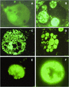
Fluorescence microscopy analysis. (A) A. castellanii trophozoite without intracellular F. tularensis (day 0). (B) Intact A. castellanii with Francisella-filled vacuoles (day 10). (C) Disintegrating A. castellanii filled with F. tularensis LVS/GFP/ASV (day 15). (D) Francisella-filled vesicles, enclosed within the cell membrane of a dead A. castellanii trophozoite (day 18). (E) Francisella-filled vesicle (day 18). (F) A. castellanii cyst containing F. tularensis LVS/GFP/ASV inside the double wall (day 40).
FIG. 3.
FIG. 3.
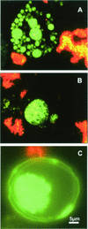
Differentiation between extracellular and intracellular F. tularensis by use of monoclonal antibodies. Viable intracellular F. tularensis LVS/GFP/AVS cells expressing GFP appear green, while extracellular F. tularensis cells appear red after treatment with labeled antibodies specific for F. tularensis. (A) Disintegrating A. castellanii trophozoite containing Francisella-filled vesicles (day 18). (B) Individual vesicle containing viable F. tularensis GFP/LVS/ASV (day 18). (C) A. castellanii cyst containing viable F. tularensis inside the cyst double wall (day 40).
FIG. 4.
FIG. 4.
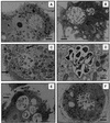
Electron microscopy analysis. (A) A. castellanii trophozoite without intracellular F. tularensis (day 0). (B) A. castellanii trophozoite with Francisella-filled vacuoles (day 9). (C and D) Recruitment of mitochondria (short arrows) and rough endoplasmic reticulum (long arrows) to the vacuole containing bacteria. (E) A. castellanii trophozoite undergoing encystation with F. tularensis cells lined up between the two layers of the emerging double wall (day 16). (F) A. castellanii cyst containing F. tularensis on the inside of the double wall (day 16).
FIG. 5.
FIG. 5.
Viable counts of F. tularensis cultured alone (squares), F. tularensis cocultured with A. castellanii (triangles), and intracellular F. tularensis (circles). Mean values from two independent experiments are shown (t test, P < 0.05). Bars show averages of the absolute deviations of data points from their mean.
FIG. 6.
FIG. 6.
Counts of A. castellanii cultured with F. tularensis (triangles) and without F. tularensis (squares) over time. Mean values from two independent experiments are shown (t test, P < 0.05). Bars show averages of the absolute deviations of data points from their mean.

References

    1. Abu Kwaik, Y., L. Y. Gao, B. J. Stone, C. Venkataraman, and O. S. Harb. 1998. Invasion of protozoa by Legionella pneumophila and its role in bacterial ecology and pathogenesis. Appl. Environ. Microbiol. 64:3127-3133. - PMC - PubMed
    1. Amann, R., N. Springer, W. Schonhuber, W. Ludwig, E. N. Schmid, K. D. Muller, and R. Michel. 1997. Obligate intracellular bacterial parasites of Acanthamoeba related to Chlamydia spp. Appl. Environ. Microbiol. 63:115-121. - PMC - PubMed
    1. Anthony, L. S. D., R. D. Burke, and F. E. Nano. 1991. Growth of Francisella spp. in rodent macrophages. Infect. Immun. 59:3291-3296. - PMC - PubMed
    1. Anthony, L. S. D., and P. A. L. Kongshavn. 1985. Host resistance to experimental murine tularemia. Fed. Proc. 44:528. - PMC - PubMed
    1. Berdal, B. P., R. Mehl, N. K. Meidell, A. Lorentzen-Styr, and O. Scheel. 1996. Field investigations of tularaemia in Norway. FEMS Immunol. Med. Microbiol. 13:191-195. - PubMed

Publication types

MeSH terms

LinkOut - more resources